4DHG

Crystal structure of enolase TBIS_1083(TARGET EFI-502310) from Thermobispora bispora dsm 43833, an open loop conformation


Experimental Data Snapshot

  • Method: X-RAY DIFFRACTION
  • Resolution: 1.90 Å
  • R-Value Free: 0.200 
  • R-Value Work: 0.161 
  • R-Value Observed: 0.162 

wwPDB Validation   3D Report Full Report


This is version 1.1 of the entry. See complete history


Literature

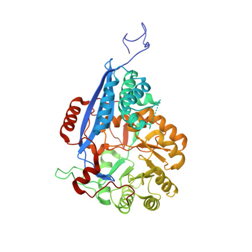
Crystal Structure of Enolase Tbis_1083 from Thermobispora Bispora Dsm 43833

Patskovsky, Y.Toro, R.Bhosle, R.Hillerich, B.Seidel, R.D.Washington, E.Scott Glenn, A.Chowdhury, S.Evans, B.Hammonds, J.Zencheck, W.D.Imker, H.J.Gerlt, J.A.Almo, S.C.

To be published.

Macromolecules
Find similar proteins by:  (by identity cutoff)  |  3D Structure
Entity ID: 1
MoleculeChains Sequence LengthOrganismDetailsImage
Mandelate racemase/muconate lactonizing protein
A, B, C, D
441Thermobispora bispora DSM 43833Mutation(s): 0 
Gene Names: Tbis_1083
UniProt
Find proteins for D6Y7Y6 (Thermobispora bispora (strain ATCC 19993 / DSM 43833 / CBS 139.67 / JCM 10125 / KCTC 9307 / NBRC 14880 / R51))
Explore D6Y7Y6 
Go to UniProtKB:  D6Y7Y6
Entity Groups  
Sequence Clusters30% Identity50% Identity70% Identity90% Identity95% Identity100% Identity
UniProt GroupD6Y7Y6
Sequence Annotations
Expand
  • Reference Sequence
Small Molecules
Ligands 4 Unique
IDChains Name / Formula / InChI Key2D Diagram3D Interactions
IOD
Query on IOD

Download Ideal Coordinates CCD File 
F [auth A]
H [auth A]
J [auth B]
M [auth B]
Q [auth D]
F [auth A],
H [auth A],
J [auth B],
M [auth B],
Q [auth D],
R [auth D],
S [auth D],
T [auth D],
U [auth D],
V [auth D],
W [auth D]
IODIDE ION
I
XMBWDFGMSWQBCA-UHFFFAOYSA-M
PO4
Query on PO4

Download Ideal Coordinates CCD File 
E [auth A],
I [auth B],
N [auth C],
P [auth D]
PHOSPHATE ION
O4 P
NBIIXXVUZAFLBC-UHFFFAOYSA-K
GOL
Query on GOL

Download Ideal Coordinates CCD File 
G [auth A],
L [auth B],
O [auth C]
GLYCEROL
C3 H8 O3
PEDCQBHIVMGVHV-UHFFFAOYSA-N
UNL
Query on UNL

Download Ideal Coordinates CCD File 
K [auth B]Unknown ligand
PEDCQBHIVMGVHV-UHFFFAOYSA-N
Experimental Data & Validation

Experimental Data

  • Method: X-RAY DIFFRACTION
  • Resolution: 1.90 Å
  • R-Value Free: 0.200 
  • R-Value Work: 0.161 
  • R-Value Observed: 0.162 
  • Space Group: P 1
Unit Cell:
Length ( Å )Angle ( ˚ )
a = 70.028α = 89.26
b = 84.823β = 84.07
c = 94.119γ = 72.72
Software Package:
Software NamePurpose
PHASERphasing
REFMACrefinement
HKL-3000data reduction
HKL-3000data scaling

Structure Validation

View Full Validation Report


Entry History 

Revision History  (Full details and data files)

  • Version 1.0: 2012-02-15
    Type: Initial release
  • Version 1.1: 2023-09-13
    Changes: Data collection, Database references, Derived calculations, Refinement description