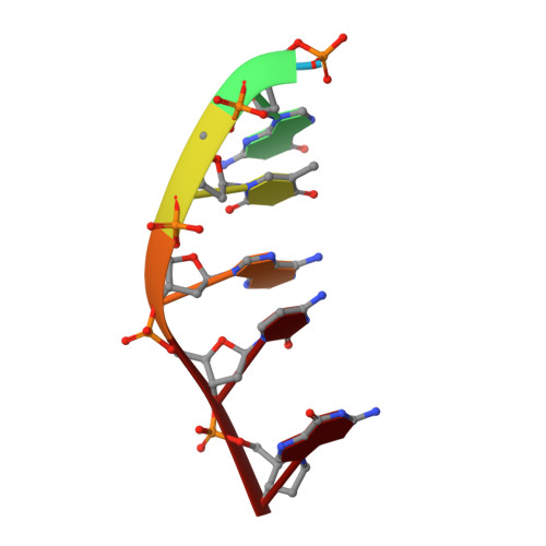
The crystal structure of Chelerythrine bound to DNA d(CGTACG)
Ferraroni, M., Bazzicalupi, C., Gratteri, P., Bilia, A.R.To be published.
Experimental Data Snapshot
| Ligands 2 Unique | |||||
|---|---|---|---|---|---|
| ID | Chains | Name / Formula / InChI Key | 2D Diagram | 3D Interactions | |
| CTI Query on CTI | E [auth A] | 1,2-dimethoxy-12-methyl[1,3]benzodioxolo[5,6-c]phenanthridin-12-ium C21 H18 N O4 LLEJIEBFSOEYIV-UHFFFAOYSA-N |  | ||
| CA Query on CA | F [auth B] | CALCIUM ION Ca BHPQYMZQTOCNFJ-UHFFFAOYSA-N |  | ||
| Length ( Å ) | Angle ( ˚ ) |
|---|---|
| a = 30.231 | α = 90 |
| b = 30.231 | β = 90 |
| c = 119.247 | γ = 120 |
| Software Name | Purpose |
|---|---|
| XSCALE | data scaling |
| MOLREP | phasing |
| SHELX | refinement |
| PDB_EXTRACT | data extraction |
| XDS | data reduction |
| XDS | data scaling |
RCSB PDB (citation) is hosted by
RCSB PDB is a member of the