4D7O

Structure of rat neuronal nitric oxide synthase heme domain in complex with 6-(4-(((3-Fluorophenethyl)amino)methyl)phenyl)-4- methylpyridin-2-amine


Experimental Data Snapshot

  • Method: X-RAY DIFFRACTION
  • Resolution: 1.78 Å
  • R-Value Free: 0.209 
  • R-Value Work: 0.179 
  • R-Value Observed: 0.181 

wwPDB Validation   3D Report Full Report


Ligand Structure Quality Assessment 


This is version 1.0 of the entry. See complete history


Literature

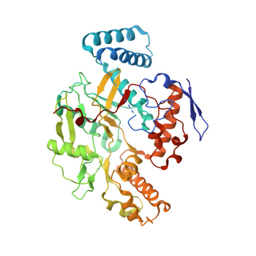
Nitric Oxide Synthase as a Target for Methicillin-Resistant Staphylococcus Aureus

Holden, J.K.Kang, S.Beasley, F.C.Cinelli, M.A.Li, H.Roy, S.G.Dejam, D.Edinger, A.L.Nizet, V.Silverman, R.B.Poulos, T.L.

(2015) Chem Biol 22: 785

  • DOI: https://doi.org/10.1016/j.chembiol.2015.05.013
  • Primary Citation of Related Structures:  
    4D7H, 4D7I, 4D7J, 4D7O

  • PubMed Abstract: 

    Bacterial infections associated with methicillin-resistant Staphylococcus aureus (MRSA) are a major economic burden to hospitals, and confer high rates of morbidity and mortality among those infected. Exploitation of novel therapeutic targets is thus necessary to combat this dangerous pathogen. Here, we report on the identification and characterization, including crystal structures, of two nitric oxide synthase (NOS) inhibitors that function as antimicrobials against MRSA. These data provide the first evidence that bacterial NOS (bNOS) inhibitors can work synergistically with oxidative stress to enhance MRSA killing. Crystal structures show that each inhibitor contacts an active site Ile residue in bNOS that is Val in the mammalian NOS isoforms. Mutagenesis studies show that the additional nonpolar contacts provided by the Ile in bNOS contribute to tighter binding toward the bacterial enzyme.


  • Organizational Affiliation

    Departments of Molecular Biology and Biochemistry, Pharmaceutical Sciences, and Chemistry, University of California, Irvine, CA 92697-3900, USA.


Macromolecules
Find similar proteins by:  (by identity cutoff)  |  3D Structure
Entity ID: 1
MoleculeChains Sequence LengthOrganismDetailsImage
NITRIC OXIDE SYNTHASE, BRAIN
A, B
422Rattus norvegicusMutation(s): 0 
EC: 1.14.13.39
UniProt
Find proteins for P29476 (Rattus norvegicus)
Explore P29476 
Go to UniProtKB:  P29476
Entity Groups  
Sequence Clusters30% Identity50% Identity70% Identity90% Identity95% Identity100% Identity
UniProt GroupP29476
Sequence Annotations
Expand
  • Reference Sequence
Small Molecules
Ligands 5 Unique
IDChains Name / Formula / InChI Key2D Diagram3D Interactions
HEM
Query on HEM

Download Ideal Coordinates CCD File 
C [auth A],
G [auth B]
PROTOPORPHYRIN IX CONTAINING FE
C34 H32 Fe N4 O4
KABFMIBPWCXCRK-RGGAHWMASA-L
0GD
Query on 0GD

Download Ideal Coordinates CCD File 
E [auth A],
I [auth B]
6-[4-({[2-(3-fluorophenyl)ethyl]amino}methyl)phenyl]-4-methylpyridin-2-amine
C21 H22 F N3
YIRCIVHQWVHKOD-UHFFFAOYSA-N
H4B
Query on H4B

Download Ideal Coordinates CCD File 
D [auth A],
H [auth B]
5,6,7,8-TETRAHYDROBIOPTERIN
C9 H15 N5 O3
FNKQXYHWGSIFBK-RPDRRWSUSA-N
ZN
Query on ZN

Download Ideal Coordinates CCD File 
K [auth B]ZINC ION
Zn
PTFCDOFLOPIGGS-UHFFFAOYSA-N
ACT
Query on ACT

Download Ideal Coordinates CCD File 
F [auth A],
J [auth B]
ACETATE ION
C2 H3 O2
QTBSBXVTEAMEQO-UHFFFAOYSA-M
Binding Affinity Annotations 
IDSourceBinding Affinity
0GD Binding MOAD:  4D7O Ki: 525 (nM) from 1 assay(s)
Experimental Data & Validation

Experimental Data

  • Method: X-RAY DIFFRACTION
  • Resolution: 1.78 Å
  • R-Value Free: 0.209 
  • R-Value Work: 0.179 
  • R-Value Observed: 0.181 
  • Space Group: P 21 21 21
Unit Cell:
Length ( Å )Angle ( ˚ )
a = 51.77α = 90
b = 110.6β = 90
c = 165.162γ = 90
Software Package:
Software NamePurpose
REFMACrefinement
HKL-2000data reduction
SCALEPACKdata scaling
REFMACphasing

Structure Validation

View Full Validation Report


Ligand Structure Quality Assessment 


Entry History 

Deposition Data

Revision History  (Full details and data files)

  • Version 1.0: 2015-07-01
    Type: Initial release