4D1X

CDK2 in complex with Luciferin


Experimental Data Snapshot

  • Method: X-RAY DIFFRACTION
  • Resolution: 2.10 Å
  • R-Value Free: 0.238 
  • R-Value Work: 0.194 
  • R-Value Observed: 0.196 

wwPDB Validation   3D Report Full Report


Ligand Structure Quality Assessment 


This is version 1.2 of the entry. See complete history


Literature

Luciferin and Derivatives as a Dyrk Selective Scaffold for the Design of Protein Kinase Inhibitors.

Rothweiler, U.Eriksson, J.Stensen, W.Leeson, F.Engh, R.A.Svendsen, J.S.

(2015) Eur J Med Chem 94: 140

  • DOI: https://doi.org/10.1016/j.ejmech.2015.02.035
  • Primary Citation of Related Structures:  
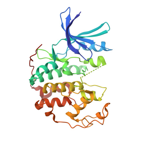
    4D1X, 4D1Z

  • PubMed Abstract: 

    D-Luciferin is widely used as a substrate in luciferase catalysed bioluminescence assays for in vitro studies. However, little is known about cross reactivity and potential interference of D-luciferin with other enzymes. We serendipitously found that firefly luciferin inhibited the CDK2/Cyclin A protein kinase. Inhibition profiling of D-luciferin over a 103-protein kinase panel showed significant inhibition of a small set of protein kinases, in particular the DYRK-family, but also other members of the CMGC-group, including ERK8 and CK2. Inhibition profiling on a 16-member focused library derived from D-luciferin confirms that D-luciferin represents a DYRK-selective chemotype of fragment-like molecular weight. Thus, observation of its inhibitory activity and the initial SAR information reported here promise to be useful for further design of protein kinase inhibitors with related scaffolds.


  • Organizational Affiliation

    The Norwegian Structural Biology Centre, Department of Chemistry, UiT The Arctic University of Norway, N-9037 Tromsø, Norway.


Macromolecules
Find similar proteins by:  (by identity cutoff)  |  3D Structure
Entity ID: 1
MoleculeChains Sequence LengthOrganismDetailsImage
CYCLIN-DEPENDENT KINASE 2298Homo sapiensMutation(s): 0 
EC: 2.7.11.22
UniProt & NIH Common Fund Data Resources
Find proteins for P24941 (Homo sapiens)
Explore P24941 
Go to UniProtKB:  P24941
PHAROS:  P24941
GTEx:  ENSG00000123374 
Entity Groups  
Sequence Clusters30% Identity50% Identity70% Identity90% Identity95% Identity100% Identity
UniProt GroupP24941
Sequence Annotations
Expand
  • Reference Sequence
Small Molecules
Ligands 1 Unique
IDChains Name / Formula / InChI Key2D Diagram3D Interactions
ESJ
Query on ESJ

Download Ideal Coordinates CCD File 
B [auth A](4S)-2-(6-hydroxy-1,3-benzothiazol-2-yl)-4,5-dihydro-1,3-thiazole-4-carboxylic acid
C11 H8 N2 O3 S2
BJGNCJDXODQBOB-SSDOTTSWSA-N
Experimental Data & Validation

Experimental Data

  • Method: X-RAY DIFFRACTION
  • Resolution: 2.10 Å
  • R-Value Free: 0.238 
  • R-Value Work: 0.194 
  • R-Value Observed: 0.196 
  • Space Group: P 21 21 21
Unit Cell:
Length ( Å )Angle ( ˚ )
a = 53.31α = 90
b = 72.12β = 90
c = 72.29γ = 90
Software Package:
Software NamePurpose
PHENIXrefinement
XDSdata reduction
XDSdata scaling
MOLREPphasing

Structure Validation

View Full Validation Report


Ligand Structure Quality Assessment 


Entry History 

Deposition Data

Revision History  (Full details and data files)

  • Version 1.0: 2015-03-18
    Type: Initial release
  • Version 1.1: 2015-03-25
    Changes: Database references
  • Version 1.2: 2023-12-20
    Changes: Data collection, Database references, Derived calculations, Other, Refinement description