


4COF
Crystal structure of a human gamma-aminobutyric acid receptor, the GABA(A)R-beta3 homopentamer
- PDB DOI: https://doi.org/10.2210/pdb4COF/pdb
- Classification: TRANSPORT PROTEIN
- Organism(s): Homo sapiens
- Expression System: Homo sapiens
- Mutation(s): No
- Membrane Protein: Yes OPMPDBTMMemProtMDmpstruc
- Deposited: 2014-01-28 Released: 2014-06-04
Experimental Data Snapshot
- Method: X-RAY DIFFRACTION
- Resolution: 2.97 Å
- R-Value Free: 0.226
- R-Value Work: 0.205
- R-Value Observed: 0.206

This is version 2.1 of the entry. See complete history.

Macromolecules
Find similar proteins by:
(by identity cutoff) | 3D Structure
Entity ID: 1 | |||||
|---|---|---|---|---|---|
| Molecule | Chains | Sequence Length | Organism | Details | Image |
| GAMMA-AMINOBUTYRIC ACID RECEPTOR SUBUNIT BETA-3 | 355 | Homo sapiens | Mutation(s): 0 Membrane Entity: Yes | 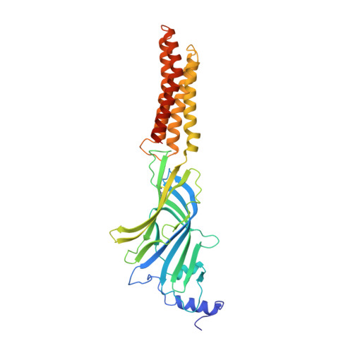 | |
UniProt & NIH Common Fund Data Resources | |||||
Find proteins for P28472 (Homo sapiens) Explore P28472 Go to UniProtKB: P28472 | |||||
PHAROS: P28472 GTEx: ENSG00000166206 | |||||
Entity Groups | |||||
| Sequence Clusters | 30% Identity50% Identity70% Identity90% Identity95% Identity100% Identity | ||||
| UniProt Group | P28472 | ||||
Sequence AnnotationsExpand | |||||
| |||||
Oligosaccharides

Small Molecules
| Ligands 3 Unique | |||||
|---|---|---|---|---|---|
| ID | Chains | Name / Formula / InChI Key | 2D Diagram | 3D Interactions | |
| NAG Query on NAG | L [auth A] N [auth A] Q [auth B] T [auth C] W [auth D] | 2-acetamido-2-deoxy-beta-D-glucopyranose C8 H15 N O6 OVRNDRQMDRJTHS-FMDGEEDCSA-N |  | ||
| BEN Query on BEN | K [auth A], O [auth B], R [auth C], U [auth D], X [auth E] | BENZAMIDINE C7 H8 N2 PXXJHWLDUBFPOL-UHFFFAOYSA-N |  | ||
| CL Query on CL | M [auth A], P [auth B], S [auth C], V [auth D], Y [auth E] | CHLORIDE ION Cl VEXZGXHMUGYJMC-UHFFFAOYSA-M |  | ||
Experimental Data & Validation
Experimental Data
- Method: X-RAY DIFFRACTION
- Resolution: 2.97 Å
- R-Value Free: 0.226
- R-Value Work: 0.205
- R-Value Observed: 0.206
- Space Group: C 1 2 1
Unit Cell:
| Length ( Å ) | Angle ( ˚ ) |
|---|---|
| a = 174.1 | α = 90 |
| b = 108.9 | β = 107.43 |
| c = 207.44 | γ = 90 |
| Software Name | Purpose |
|---|---|
| BUSTER | refinement |
| XDS | data reduction |
| SCALA | data scaling |
| PHASER | phasing |
Entry History
Deposition Data
- Released Date: 2014-06-04 Deposition Author(s): Miller, P.S., Aricescu, A.R.
Revision History (Full details and data files)
- Version 1.0: 2014-06-04
Type: Initial release - Version 1.1: 2014-06-11
Changes: Structure summary - Version 1.2: 2014-06-18
Changes: Database references - Version 1.3: 2014-08-27
Changes: Database references - Version 2.0: 2020-07-29
Type: Remediation
Reason: Carbohydrate remediation
Changes: Atomic model, Data collection, Derived calculations, Other, Structure summary - Version 2.1: 2023-12-20
Changes: Data collection, Database references, Derived calculations, Refinement description, Structure summary
