4CII

Crystal structure of N-terminally truncated Helicobacter pylori T4SS Protein CagL as domain swapped dimer


Experimental Data Snapshot

  • Method: X-RAY DIFFRACTION
  • Resolution: 2.15 Å
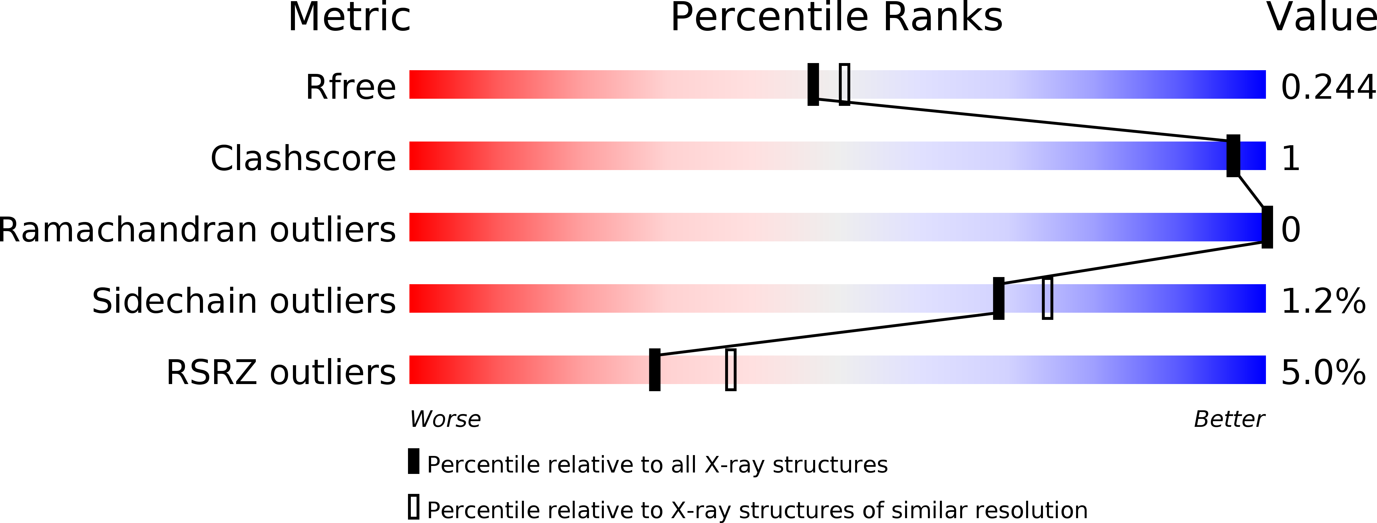
  • R-Value Free: 0.237 
  • R-Value Work: 0.199 
  • R-Value Observed: 0.200 

wwPDB Validation   3D Report Full Report


This is version 1.4 of the entry. See complete history


Literature

Structure of a Three-Dimensional Domain-Swapped Dimer of the Helicobacter Pylori Type Iv Secretion System Pilus Protein Cagl.

Barden, S.Schomburg, B.Conradi, J.Backert, S.Sewald, N.Niemann, H.H.

(2014) Acta Crystallogr D Biol Crystallogr 70: 1391

  • DOI: https://doi.org/10.1107/S1399004714003150
  • Primary Citation of Related Structures:  
    4CII

  • PubMed Abstract: 

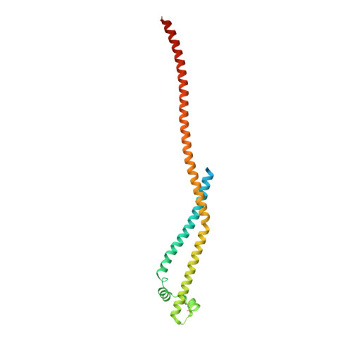
    A new crystal form of the Helicobacter pylori type IV secretion system (T4SS) pilus protein CagL is described here. In contrast to two previously reported monomeric structures, CagL forms a three-dimensional domain-swapped dimer. CagL dimers can arise during refolding from inclusion bodies or can form spontaneously from purified monomeric CagL in the crystallization conditions. Monomeric CagL forms a three-helix bundle, with which the N-terminal helix is only loosely associated. In the new crystal form, the N-terminal helix is missing. The domain swap is owing to exchange of the C-terminal helix between the two protomers of a dimer. A loop-to-helix transition results in a long helix of 108 amino acids comprising the penultimate and the last helix of the monomer. The RGD motif of dimeric CagL adopts an α-helical conformation. In contrast to the previously reported structures, the conserved and functionally important C-terminal hexapeptide is resolved. It extends beyond the three-helix bundle as an exposed helical appendage. This new crystal form contributes to the molecular understanding of CagL by highlighting rigid and flexible regions in the protein and by providing the first view of the C-terminus. Based on the structural features, a previously unrecognized homology between CagL and CagI is discussed.


  • Organizational Affiliation

    Structural Biochemistry, Department of Chemistry, Bielefeld University, Universitätsstrasse 25, 33615 Bielefeld, Germany.


Macromolecules
Find similar proteins by:  (by identity cutoff)  |  3D Structure
Entity ID: 1
MoleculeChains Sequence LengthOrganismDetailsImage
CAG PATHOGENICITY ISLAND PROTEIN (CAG18)225Helicobacter pylori 26695Mutation(s): 0 
UniProt
Find proteins for O25272 (Helicobacter pylori (strain ATCC 700392 / 26695))
Explore O25272 
Go to UniProtKB:  O25272
Entity Groups  
Sequence Clusters30% Identity50% Identity70% Identity90% Identity95% Identity100% Identity
UniProt GroupO25272
Sequence Annotations
Expand
  • Reference Sequence
Small Molecules
Ligands 1 Unique
IDChains Name / Formula / InChI Key2D Diagram3D Interactions
MRD
Query on MRD

Download Ideal Coordinates CCD File 
B [auth A](4R)-2-METHYLPENTANE-2,4-DIOL
C6 H14 O2
SVTBMSDMJJWYQN-RXMQYKEDSA-N
Experimental Data & Validation

Experimental Data

  • Method: X-RAY DIFFRACTION
  • Resolution: 2.15 Å
  • R-Value Free: 0.237 
  • R-Value Work: 0.199 
  • R-Value Observed: 0.200 
  • Space Group: P 65 2 2
Unit Cell:
Length ( Å )Angle ( ˚ )
a = 61.841α = 90
b = 61.841β = 90
c = 244.499γ = 120
Software Package:
Software NamePurpose
PHENIXrefinement
XDSdata reduction
SCALAdata scaling
PHASERphasing

Structure Validation

View Full Validation Report


Entry History 

Deposition Data

Revision History  (Full details and data files)

  • Version 1.0: 2014-02-19
    Type: Initial release
  • Version 1.1: 2014-05-14
    Changes: Database references
  • Version 1.2: 2014-05-21
    Changes: Database references
  • Version 1.3: 2019-03-06
    Changes: Data collection, Experimental preparation, Other
  • Version 1.4: 2019-09-25
    Changes: Data collection, Experimental preparation, Other