4BQA

Crystal structure of the ETS domain of human ETS2 in complex with DNA


Experimental Data Snapshot

  • Method: X-RAY DIFFRACTION
  • Resolution: 2.50 Å
  • R-Value Free: 0.255 
  • R-Value Work: 0.234 
  • R-Value Observed: 0.235 

wwPDB Validation   3D Report Full Report


This is version 1.3 of the entry. See complete history


Literature

Structural Insights Into the Autoregulation and Cooperativity of the Human Transcription Factor Ets-2.

Newman, J.A.Cooper, C.D.O.Aitkenhead, H.Gileadi, O.

(2015) J Biol Chem 290: 8539

  • DOI: https://doi.org/10.1074/jbc.M114.619270
  • Primary Citation of Related Structures:  
    4BQA

  • PubMed Abstract: 

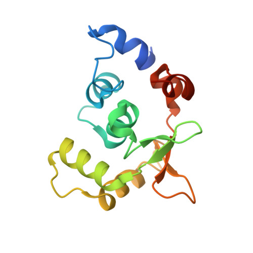
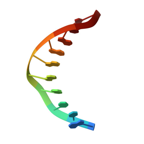
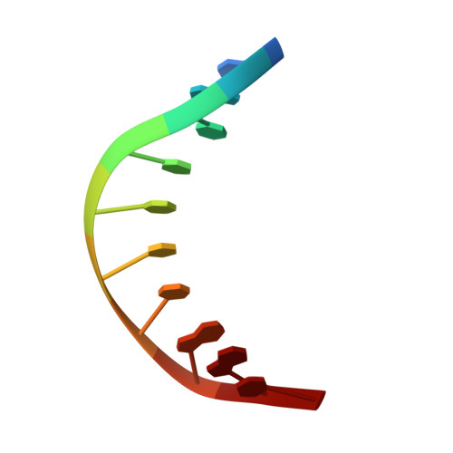
    Ets-2, like its closely related homologue Ets-1, is a member of the Ets family of DNA binding transcription factors. Both proteins are subject to multiple levels of regulation of their DNA binding and transactivation properties. One such regulatory mechanism is the presence of an autoinhibitory module, which in Ets-1 allosterically inhibits the DNA binding activity. This inhibition can be relieved by interaction with protein partners or cooperative binding to closely separated Ets binding sites in a palindromic arrangement. In this study we describe the 2.5 Å resolution crystal structure of a DNA complex of the Ets-2 Ets domain. The Ets domain crystallized with two distinct species in the asymmetric unit, which closely resemble the autoinhibited and DNA bound forms of Ets-1. This discovery prompted us to re-evaluate the current model for the autoinhibitory mechanism and the structural basis for cooperative DNA binding. In contrast to Ets-1, in which the autoinhibition is caused by a combination of allosteric and steric mechanisms, we were unable to find clear evidence for the allosteric mechanism in Ets-2. We also demonstrated two possibly distinct types of cooperative binding to substrates with Ets binding motifs separated by four and six base pairs and suggest possible molecular mechanisms for this behavior.


  • Organizational Affiliation

    From the Structural Genomics Consortium, University of Oxford, Oxford OX3 7DQ, United Kingdom.


Macromolecules

Find similar proteins by:  (by identity cutoff)  |  3D Structure
Entity ID: 1
MoleculeChains Sequence LengthOrganismDetailsImage
PROTEIN C-ETS-2
A, D, G
142Homo sapiensMutation(s): 0 
UniProt & NIH Common Fund Data Resources
Find proteins for P15036 (Homo sapiens)
Explore P15036 
Go to UniProtKB:  P15036
PHAROS:  P15036
GTEx:  ENSG00000157557 
Entity Groups  
Sequence Clusters30% Identity50% Identity70% Identity90% Identity95% Identity100% Identity
UniProt GroupP15036
Sequence Annotations
Expand
  • Reference Sequence

Find similar nucleic acids by:  Sequence   |   3D Structure  

Entity ID: 2
MoleculeChains LengthOrganismImage
5'-D(*AP*CP*CP*GP*GP*AP*AP*GP*TP*GP)-3'
B, E, H
10Homo sapiens
Sequence Annotations
Expand
  • Reference Sequence

Find similar nucleic acids by:  Sequence   |   3D Structure  

Entity ID: 3
MoleculeChains LengthOrganismImage
5'-D(*CP*AP*CP*TP*TP*CP*CP*GP*GP*TP)-3'
C, F, I
10Homo sapiens
Sequence Annotations
Expand
  • Reference Sequence
Experimental Data & Validation

Experimental Data

  • Method: X-RAY DIFFRACTION
  • Resolution: 2.50 Å
  • R-Value Free: 0.255 
  • R-Value Work: 0.234 
  • R-Value Observed: 0.235 
  • Space Group: P 1 21 1
Unit Cell:
Length ( Å )Angle ( ˚ )
a = 36.674α = 90
b = 96.964β = 97.06
c = 83.803γ = 90
Software Package:
Software NamePurpose
PHENIXrefinement
XDSdata reduction
XDSdata scaling
MOLREPphasing

Structure Validation

View Full Validation Report


Entry History 

Deposition Data

Revision History  (Full details and data files)

  • Version 1.0: 2013-06-12
    Type: Initial release
  • Version 1.1: 2015-04-01
    Changes: Database references
  • Version 1.2: 2015-04-15
    Changes: Database references
  • Version 1.3: 2023-12-20
    Changes: Data collection, Database references, Other, Refinement description