4B5S

Crystal structures of divalent metal dependent pyruvate aldolase, HpaI, in complex with pyruvate


Experimental Data Snapshot

  • Method: X-RAY DIFFRACTION
  • Resolution: 1.68 Å
  • R-Value Free: 0.179 
  • R-Value Work: 0.151 
  • R-Value Observed: 0.154 

wwPDB Validation   3D Report Full Report


This is version 3.1 of the entry. See complete history


Literature

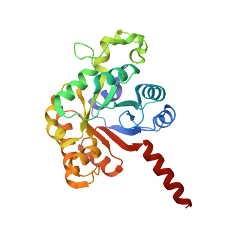
Crystal Structure of Reaction Intermediates in Pyruvate Class II Aldolase: Substrate Cleavage, Enolate Stabilization and Substrate Specificity

Coincon, M.Wang, W.Sygusch, J.Seah, S.Y.K.

(2012) J Biol Chem 287: 36208

  • DOI: https://doi.org/10.1074/jbc.M112.400705
  • Primary Citation of Related Structures:  
    4B5S, 4B5T, 4B5U, 4B5V, 4B5W, 4B5X

  • PubMed Abstract: 

    Crystal structures of divalent metal-dependent pyruvate aldolase, HpaI, in complex with substrate and cleavage products were determined to 1.8-2.0 Å resolution. The enzyme·substrate complex with 4-hydroxy-2-ketoheptane-1,7-dioate indicates that water molecule W2 bound to the divalent metal ion initiates C3-C4 bond cleavage. The binding mode of the aldehyde donor delineated a solvent-filled capacious binding locus lined with predominantly hydrophobic residues. The absence of direct interactions with the aldehyde aliphatic carbons accounts for the broad specificity and lack of stereospecific control by the enzyme. Enzymatic complex structures formed with keto acceptors, pyruvate, and 2-ketobutyrate revealed bidentate interaction with the divalent metal ion by C1-carboxyl and C2-carbonyl oxygens and water molecule W4 that is within close contact of the C3 carbon. Arg(70) assumes a multivalent role through its guanidinium moiety interacting with all active site enzymatic species: C2 oxygen in substrate, pyruvate, and ketobutyrate; substrate C4 hydroxyl; aldehyde C1 oxygen; and W4. The multiple interactions made by Arg(70) stabilize the negatively charged C4 oxygen following proton abstraction, the aldehyde alignment in aldol condensation, and the pyruvate enolate upon aldol cleavage as well as support proton exchange at C3. This role is corroborated by loss of aldol cleavage ability and pyruvate C3 proton exchange activity and by a 730-fold increase in the dissociation constant toward the pyruvate enolate analog oxalate in the R70A mutant. Based on the crystal structures, a mechanism is proposed involving the two enzyme-bound water molecules, W2 and W4, in acid/base catalysis that facilitates reversible aldol cleavage. The same reaction mechanism promotes decarboxylation of oxaloacetate.


  • Organizational Affiliation

    Department of Biochemistry, Université de Montréal, Montréal, Québec H3C 3J7, Canada.


Macromolecules
Find similar proteins by:  (by identity cutoff)  |  3D Structure
Entity ID: 1
MoleculeChains Sequence LengthOrganismDetailsImage
4-HYDROXY-2-OXO-HEPTANE-1,7-DIOATE ALDOLASE
A, B
251Escherichia coli ATCC 8739Mutation(s): 0 
EC: 4.1.2.20
UniProt
Find proteins for B1IS70 (Escherichia coli (strain ATCC 8739 / DSM 1576 / NBRC 3972 / NCIMB 8545 / WDCM 00012 / Crooks))
Explore B1IS70 
Go to UniProtKB:  B1IS70
Entity Groups  
Sequence Clusters30% Identity50% Identity70% Identity90% Identity95% Identity100% Identity
UniProt GroupB1IS70
Sequence Annotations
Expand
  • Reference Sequence
Small Molecules
Ligands 6 Unique
IDChains Name / Formula / InChI Key2D Diagram3D Interactions
PO4
Query on PO4

Download Ideal Coordinates CCD File 
E [auth A],
F [auth A],
K [auth B],
L [auth B]
PHOSPHATE ION
O4 P
NBIIXXVUZAFLBC-UHFFFAOYSA-K
GOL
Query on GOL

Download Ideal Coordinates CCD File 
I [auth B]GLYCEROL
C3 H8 O3
PEDCQBHIVMGVHV-UHFFFAOYSA-N
3GR
Query on 3GR

Download Ideal Coordinates CCD File 
C [auth A]D-Glyceraldehyde
C3 H6 O3
MNQZXJOMYWMBOU-VKHMYHEASA-N
PYR
Query on PYR

Download Ideal Coordinates CCD File 
G [auth A],
M [auth B]
PYRUVIC ACID
C3 H4 O3
LCTONWCANYUPML-UHFFFAOYSA-N
CO
Query on CO

Download Ideal Coordinates CCD File 
D [auth A],
J [auth B]
COBALT (II) ION
Co
XLJKHNWPARRRJB-UHFFFAOYSA-N
MG
Query on MG

Download Ideal Coordinates CCD File 
H [auth A],
N [auth B]
MAGNESIUM ION
Mg
JLVVSXFLKOJNIY-UHFFFAOYSA-N
Binding Affinity Annotations 
IDSourceBinding Affinity
PYR PDBBind:  4B5S Kd: 3.20e+5 (nM) from 1 assay(s)
Experimental Data & Validation

Experimental Data

  • Method: X-RAY DIFFRACTION
  • Resolution: 1.68 Å
  • R-Value Free: 0.179 
  • R-Value Work: 0.151 
  • R-Value Observed: 0.154 
  • Space Group: P 21 3
Unit Cell:
Length ( Å )Angle ( ˚ )
a = 151.74α = 90
b = 151.74β = 90
c = 151.74γ = 90
Software Package:
Software NamePurpose
PHENIXrefinement
PHENIXphasing

Structure Validation

View Full Validation Report


Entry History 

Deposition Data

Revision History  (Full details and data files)

  • Version 1.0: 2012-08-29
    Type: Initial release
  • Version 1.1: 2012-09-05
    Changes: Atomic model, Database references
  • Version 1.2: 2012-10-31
    Changes: Database references
  • Version 1.3: 2013-02-06
    Changes: Atomic model
  • Version 1.4: 2017-07-12
    Changes: Refinement description
  • Version 2.0: 2020-07-08
    Changes: Advisory, Atomic model, Data collection, Derived calculations, Other, Structure summary
  • Version 3.0: 2023-11-15
    Changes: Atomic model, Data collection, Database references, Derived calculations
  • Version 3.1: 2023-12-20
    Changes: Refinement description