Intersubunit Ionic Interactions Stabilize the Nucleoside Diphosphate Kinase of Mycobacterium Tuberculosis.
Georgescauld, F., Moynie, L., Habersetzer, J., Cervoni, L., Mocan, I., Borza, T., Harris, P., Dautant, A., Lascu, I.(2013) PLoS One 8: 57867
- PubMed: 23526954
- DOI: https://doi.org/10.1371/journal.pone.0057867
- Primary Citation of Related Structures:
4ANC, 4AND - PubMed Abstract:
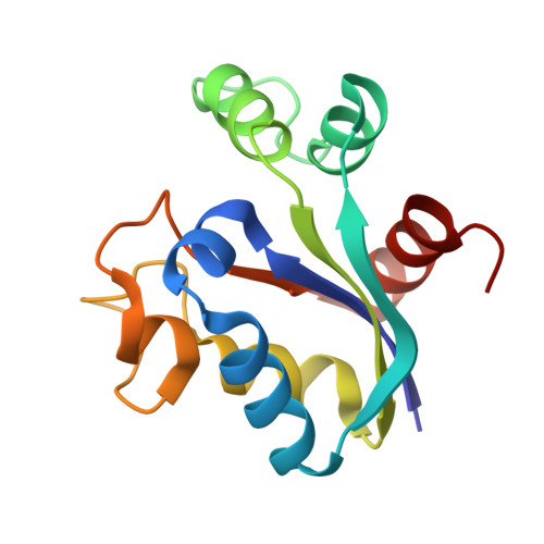
Most nucleoside diphosphate kinases (NDPKs) are hexamers. The C-terminal tail interacting with the neighboring subunits is crucial for hexamer stability. In the NDPK from Mycobacterium tuberculosis (Mt) this tail is missing. The quaternary structure of Mt-NDPK is essential for full enzymatic activity and for protein stability to thermal and chemical denaturation. We identified the intersubunit salt bridge Arg(80)-Asp(93) as essential for hexamer stability, compensating for the decreased intersubunit contact area. Breaking the salt bridge by the mutation D93N dramatically decreased protein thermal stability. The mutation also decreased stability to denaturation by urea and guanidinium. The D93N mutant was still hexameric and retained full activity. When exposed to low concentrations of urea it dissociated into folded monomers followed by unfolding while dissociation and unfolding of the wild type simultaneously occur at higher urea concentrations. The dissociation step was not observed in guanidine hydrochloride, suggesting that low concentration of salt may stabilize the hexamer. Indeed, guanidinium and many other salts stabilized the hexamer with a half maximum effect of about 0.1 M, increasing protein thermostability. The crystal structure of the D93N mutant has been solved.
Organizational Affiliation:
IBGC, University Bordeaux, Bordeaux, France.