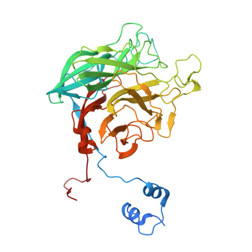
4AK7

Crystal structure of BpGH117_E303Q in complex with neoagarobiose


Experimental Data Snapshot

  • Method: X-RAY DIFFRACTION
  • Resolution: 1.80 Å
  • R-Value Free: 0.209 
  • R-Value Work: 0.155 
  • R-Value Observed: 0.158 

wwPDB Validation   3D Report Full Report


Ligand Structure Quality Assessment 


This is version 1.1 of the entry. See complete history


Literature

Analysis of Keystone Enzyme in Agar Hydrolysis Provides Insight Into the Degradation (of a Polysaccharide from) Red Seaweeds.

Hehemann, J.H.Smyth, L.Yadav, A.Vocadlo, D.J.Boraston, A.B.

(2012) J Biol Chem 287: 13985

  • DOI: https://doi.org/10.1074/jbc.M112.345645
  • Primary Citation of Related Structures:  
    4AK5, 4AK6, 4AK7

  • PubMed Abstract: 

    Agars are abundant polysaccharides from marine red algae, and their chemical structure consists of alternating D-galactose and 3,6-anhydro-L-galactose residues, the latter of which are presumed to make the polymer recalcitrant to degradation by most terrestrial bacteria. Here we study a family 117 glycoside hydrolase (BpGH117) encoded within a recently discovered locus from the human gut bacterium Bacteroides plebeius. Consistent with this locus being involved in agarocolloid degradation, we show that BpGH117 is an exo-acting 3,6-anhydro-α-(1,3)-L-galactosidase that removes the 3,6-anhydrogalactose from the non-reducing end of neoagaro-oligosaccharides. A Michaelis complex of BpGH117 with neoagarobiose reveals the distortion of the constrained 3,6-anhydro-L-galactose into a conformation that favors catalysis. Furthermore, this complex, supported by analysis of site-directed mutants, provides evidence for an organization of the active site and positioning of the catalytic residues that are consistent with an inverting mechanism of catalysis and suggests that a histidine residue acts as the general acid. This latter feature differs from the vast majority of glycoside hydrolases, which use a carboxylic acid, highlighting the alternative strategies that enzymes may utilize in catalyzing the cleavage of glycosidic bonds.


  • Organizational Affiliation

    Department of Biochemistry and Microbiology, University of Victoria, PO Box 3055 STN CSC, Victoria, British Columbia V8W 3P6, Canada.


Macromolecules
Find similar proteins by:  (by identity cutoff)  |  3D Structure
Entity ID: 1
MoleculeChains Sequence LengthOrganismDetailsImage
ANHYDRO-ALPHA-L-GALACTOSIDASE
A, B
404Phocaeicola plebeiusMutation(s): 1 
UniProt
Find proteins for B5CY74 (Phocaeicola plebeius (strain DSM 17135 / JCM 12973 / M2))
Explore B5CY74 
Go to UniProtKB:  B5CY74
Entity Groups  
Sequence Clusters30% Identity50% Identity70% Identity90% Identity95% Identity100% Identity
UniProt GroupB5CY74
Sequence Annotations
Expand
  • Reference Sequence
Small Molecules
Ligands 6 Unique
IDChains Name / Formula / InChI Key2D Diagram3D Interactions
47N
Query on 47N

Download Ideal Coordinates CCD File 
C [auth A],
I [auth B]
NEOAGAROBIOSE
C12 H20 O10
JWMBOBQNPBCYER-AHBMTURSSA-N
EDO
Query on EDO

Download Ideal Coordinates CCD File 
E [auth A],
F [auth A]
1,2-ETHANEDIOL
C2 H6 O2
LYCAIKOWRPUZTN-UHFFFAOYSA-N
ACT
Query on ACT

Download Ideal Coordinates CCD File 
G [auth A],
L [auth B]
ACETATE ION
C2 H3 O2
QTBSBXVTEAMEQO-UHFFFAOYSA-M
CA
Query on CA

Download Ideal Coordinates CCD File 
D [auth A]CALCIUM ION
Ca
BHPQYMZQTOCNFJ-UHFFFAOYSA-N
CL
Query on CL

Download Ideal Coordinates CCD File 
J [auth B]CHLORIDE ION
Cl
VEXZGXHMUGYJMC-UHFFFAOYSA-M
MG
Query on MG

Download Ideal Coordinates CCD File 
H [auth A],
K [auth B]
MAGNESIUM ION
Mg
JLVVSXFLKOJNIY-UHFFFAOYSA-N
Experimental Data & Validation

Experimental Data

  • Method: X-RAY DIFFRACTION
  • Resolution: 1.80 Å
  • R-Value Free: 0.209 
  • R-Value Work: 0.155 
  • R-Value Observed: 0.158 
  • Space Group: P 21 21 21
Unit Cell:
Length ( Å )Angle ( ˚ )
a = 83.9α = 90
b = 93.55β = 90
c = 104.96γ = 90
Software Package:
Software NamePurpose
REFMACrefinement
MOSFLMdata reduction
SCALAdata scaling
MOLREPphasing

Structure Validation

View Full Validation Report


Ligand Structure Quality Assessment 


Entry History 

Deposition Data

Revision History  (Full details and data files)

  • Version 1.0: 2012-03-14
    Type: Initial release
  • Version 1.1: 2012-05-02
    Changes: Other