


A Detailed Study of Water Network in the Minor Groove of D(Cgcgaattcgcg)2 and D(Cgcaaatttgcg)2 in Presence of Linear Groove Binders
Munnur, D.G., Mitchell, E.P., Forsyth, V.T., Teixeira, S.C.M., Neidle, S.To be published.
Experimental Data Snapshot
Find similar nucleic acids by: Sequence | 3D Structure
Entity ID: 1 | |||||
|---|---|---|---|---|---|
| Molecule | Chains | Length | Organism | Image | |
| 5'-D(*CP*GP*CP*AP*AP*AP*TP*TP*TP*GP*CP*GP)-3' | 12 | synthetic construct | 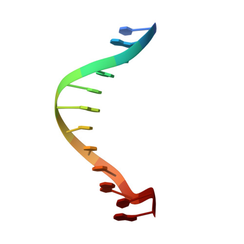 | ||
Sequence AnnotationsExpand | |||||
| |||||
| Ligands 2 Unique | |||||
|---|---|---|---|---|---|
| ID | Chains | Name / Formula / InChI Key | 2D Diagram | 3D Interactions | |
| D98 Query on D98 | F [auth B] | 2-(4'-CARBAMIMIDOYL-2'-HYDROXYBIPHENYL-4-YL)-1H-BENZIMIDAZOLE-5-CARBOXIMIDAMIDE C21 H18 N6 O VYJOWVQQJBILOA-UHFFFAOYSA-N |  | ||
| MG Query on MG | C [auth A], D [auth B], E [auth B] | MAGNESIUM ION Mg JLVVSXFLKOJNIY-UHFFFAOYSA-N |  | ||
| Length ( Å ) | Angle ( ˚ ) |
|---|---|
| a = 24.285 | α = 90 |
| b = 40.287 | β = 90 |
| c = 64.678 | γ = 90 |
| Software Name | Purpose |
|---|---|
| REFMAC | refinement |
| MOSFLM | data reduction |
| SCALA | data scaling |
| PHASER | phasing |
RCSB PDB (citation) is hosted by
RCSB PDB is a member of the

