4A8X

Structure of the core ASAP complex


Experimental Data Snapshot

  • Method: X-RAY DIFFRACTION
  • Resolution: 1.90 Å
  • R-Value Free: 0.226 
  • R-Value Work: 0.186 
  • R-Value Observed: 0.188 

wwPDB Validation   3D Report Full Report


This is version 1.2 of the entry. See complete history


Literature

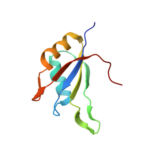
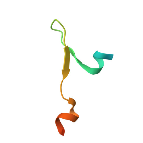
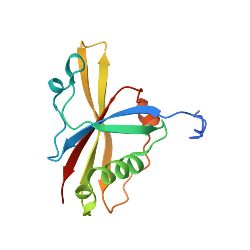
The Structure of the Asap Core Complex Reveals the Existence of a Pinin-Containing Psap Complex

Murachelli, A.G.Ebert, J.Basquin, C.Le Hir, H.Conti, E.

(2012) Nat Struct Mol Biol 19: 378

  • DOI: https://doi.org/10.1038/nsmb.2242
  • Primary Citation of Related Structures:  
    4A6Q, 4A8X, 4A90

  • PubMed Abstract: 

    The ASAP complex interacts with the exon-junction complex (EJC), a messenger ribonucleoprotein complex involved in post-transcriptional regulation. The three ASAP subunits (Acinus, RNPS1 and SAP18) have been individually implicated in transcriptional regulation, pre-mRNA splicing and mRNA quality control. To shed light on the basis for and consequences of ASAP's interaction with the EJC, we have determined the 1.9-Å resolution structure of a eukaryotic ASAP core complex. The RNA-recognition motif of RNPS1 binds to a conserved motif of Acinus with a recognition mode similar to that observed in splicing U2AF proteins. The Acinus-RNPS1 platform recruits the ubiquitin-like domain of SAP18, forming a ternary complex that has both RNA- and protein-binding properties. Unexpectedly, our structural analysis identified an Acinus-like motif in Pinin, another EJC-associated splicing factor. We show that Pinin physically interacts with RNPS1 and SAP18, forming an alternative ternary complex, PSAP.


  • Organizational Affiliation

    Max Planck Institute of Biochemistry, Department of Structural Cell Biology, Martinsried, Germany.


Macromolecules
Find similar proteins by:  (by identity cutoff)  |  3D Structure
Entity ID: 1
MoleculeChains Sequence LengthOrganismDetailsImage
RNA-BINDING PROTEIN WITH SERINE-RICH DOMAIN 188Homo sapiensMutation(s): 0 
UniProt & NIH Common Fund Data Resources
Find proteins for Q15287 (Homo sapiens)
Explore Q15287 
Go to UniProtKB:  Q15287
PHAROS:  Q15287
GTEx:  ENSG00000205937 
Entity Groups  
Sequence Clusters30% Identity50% Identity70% Identity90% Identity95% Identity100% Identity
UniProt GroupQ15287
Sequence Annotations
Expand
  • Reference Sequence
Find similar proteins by:  (by identity cutoff)  |  3D Structure
Entity ID: 2
MoleculeChains Sequence LengthOrganismDetailsImage
HOOK-LIKE, ISOFORM A40Drosophila melanogasterMutation(s): 0 
UniProt
Find proteins for Q9VJ12 (Drosophila melanogaster)
Explore Q9VJ12 
Go to UniProtKB:  Q9VJ12
Entity Groups  
Sequence Clusters30% Identity50% Identity70% Identity90% Identity95% Identity100% Identity
UniProt GroupQ9VJ12
Sequence Annotations
Expand
  • Reference Sequence
Find similar proteins by:  (by identity cutoff)  |  3D Structure
Entity ID: 3
MoleculeChains Sequence LengthOrganismDetailsImage
HISTONE DEACETYLASE COMPLEX SUBUNIT SAP18130Mus musculusMutation(s): 0 
UniProt & NIH Common Fund Data Resources
Find proteins for O55128 (Mus musculus)
Explore O55128 
Go to UniProtKB:  O55128
IMPC:  MGI:1277978
Entity Groups  
Sequence Clusters30% Identity50% Identity70% Identity90% Identity95% Identity100% Identity
UniProt GroupO55128
Sequence Annotations
Expand
  • Reference Sequence
Experimental Data & Validation

Experimental Data

  • Method: X-RAY DIFFRACTION
  • Resolution: 1.90 Å
  • R-Value Free: 0.226 
  • R-Value Work: 0.186 
  • R-Value Observed: 0.188 
  • Space Group: P 1 21 1
Unit Cell:
Length ( Å )Angle ( ˚ )
a = 38.868α = 90
b = 73.331β = 110.99
c = 50.754γ = 90
Software Package:
Software NamePurpose
PHENIXrefinement
XDSdata reduction
SCALAdata scaling
PHASERphasing

Structure Validation

View Full Validation Report


Entry History 

Deposition Data

Revision History  (Full details and data files)

  • Version 1.0: 2012-03-07
    Type: Initial release
  • Version 1.1: 2012-04-18
    Changes: Other
  • Version 1.2: 2023-12-20
    Changes: Data collection, Database references, Other, Refinement description