4A5M

Redox regulator HypR in its oxidized form


Experimental Data Snapshot

  • Method: X-RAY DIFFRACTION
  • Resolution: 3.00 Å
  • R-Value Free: 0.250 
  • R-Value Work: 0.207 
  • R-Value Observed: 0.210 

wwPDB Validation   3D Report Full Report


This is version 1.2 of the entry. See complete history


Literature

Structural Insights Into the Redox-Switch Mechanism of the Marr/Duf24-Type Regulator Hypr

Palm, G.J.Chi, B.K.Waack, P.Gronau, K.Becher, D.Albrecht, D.Hinrichs, W.Read, R.J.Antelmann, H.

(2012) Nucleic Acids Res 40: 4178

  • DOI: https://doi.org/10.1093/nar/gkr1316
  • Primary Citation of Related Structures:  
    4A5M, 4A5N

  • PubMed Abstract: 

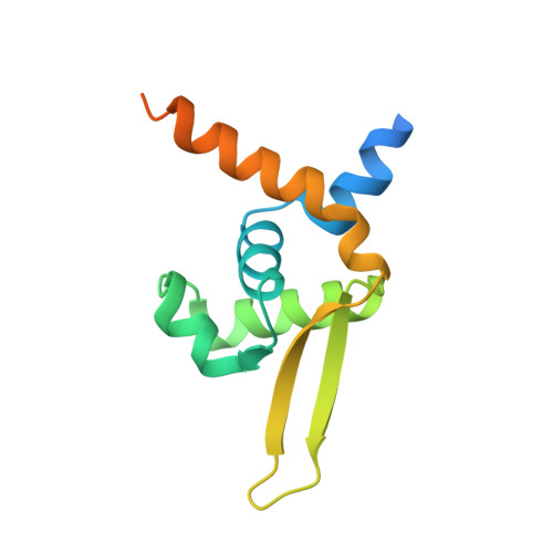
    Bacillus subtilis encodes redox-sensing MarR-type regulators of the OhrR and DUF24-families that sense organic hydroperoxides, diamide, quinones or aldehydes via thiol-based redox-switches. In this article, we characterize the novel redox-sensing MarR/DUF24-family regulator HypR (YybR) that is activated by disulphide stress caused by diamide and NaOCl in B. subtilis. HypR controls positively a flavin oxidoreductase HypO that confers protection against NaOCl stress. The conserved N-terminal Cys14 residue of HypR has a lower pK(a) of 6.36 and is essential for activation of hypO transcription by disulphide stress. HypR resembles a 2-Cys-type regulator that is activated by Cys14-Cys49' intersubunit disulphide formation. The crystal structures of reduced and oxidized HypR proteins were resolved revealing structural changes of HypR upon oxidation. In reduced HypR a hydrogen-bonding network stabilizes the reactive Cys14 thiolate that is 8-9 Å apart from Cys49'. HypR oxidation breaks these H-bonds, reorients the monomers and moves the major groove recognition α4 and α4' helices ∼4 Å towards each other. This is the first crystal structure of a redox-sensing MarR/DUF24 family protein in bacteria that is activated by NaOCl stress. Since hypochloric acid is released by activated macrophages, related HypR-like regulators could function to protect pathogens against the host immune defense.


  • Organizational Affiliation

    Institute for Biochemistry, Ernst-Moritz-Arndt-University of Greifswald, D-17487 Greifswald, Germany.


Macromolecules
Find similar proteins by:  (by identity cutoff)  |  3D Structure
Entity ID: 1
MoleculeChains Sequence LengthOrganismDetailsImage
UNCHARACTERIZED HTH-TYPE TRANSCRIPTIONAL REGULATOR YYBR
A, B, C, D, E
A, B, C, D, E, F, G, H
131Bacillus subtilisMutation(s): 0 
UniProt
Find proteins for P37486 (Bacillus subtilis (strain 168))
Explore P37486 
Go to UniProtKB:  P37486
Entity Groups  
Sequence Clusters30% Identity50% Identity70% Identity90% Identity95% Identity100% Identity
UniProt GroupP37486
Sequence Annotations
Expand
  • Reference Sequence
Small Molecules
Ligands 2 Unique
IDChains Name / Formula / InChI Key2D Diagram3D Interactions
EDO
Query on EDO

Download Ideal Coordinates CCD File 
I [auth A]
J [auth A]
K [auth A]
L [auth A]
M [auth D]
I [auth A],
J [auth A],
K [auth A],
L [auth A],
M [auth D],
N [auth E],
O [auth E],
P [auth E],
Q [auth E]
1,2-ETHANEDIOL
C2 H6 O2
LYCAIKOWRPUZTN-UHFFFAOYSA-N
CL
Query on CL

Download Ideal Coordinates CCD File 
R [auth F]CHLORIDE ION
Cl
VEXZGXHMUGYJMC-UHFFFAOYSA-M
Experimental Data & Validation

Experimental Data

  • Method: X-RAY DIFFRACTION
  • Resolution: 3.00 Å
  • R-Value Free: 0.250 
  • R-Value Work: 0.207 
  • R-Value Observed: 0.210 
  • Space Group: P 65
Unit Cell:
Length ( Å )Angle ( ˚ )
a = 82.851α = 90
b = 82.851β = 90
c = 354.436γ = 120
Software Package:
Software NamePurpose
REFMACrefinement
XDSdata reduction
XDSdata scaling
PHASERphasing

Structure Validation

View Full Validation Report


Entry History 

Deposition Data

Revision History  (Full details and data files)

  • Version 1.0: 2012-01-11
    Type: Initial release
  • Version 1.1: 2012-01-25
    Changes: Other
  • Version 1.2: 2012-05-23
    Changes: Other