3WGT

Crystal structure of D-amino acid oxidase mutant


Experimental Data Snapshot

  • Method: X-RAY DIFFRACTION
  • Resolution: 1.88 Å
  • R-Value Free: 0.230 
  • R-Value Work: 0.188 
  • R-Value Observed: 0.191 

wwPDB Validation   3D Report Full Report


Ligand Structure Quality Assessment 


This is version 1.2 of the entry. See complete history


Literature

Tailoring D-amino acid oxidase from the pig kidney to R-stereoselective amine oxidase and its use in the deracemization of alpha-methylbenzylamine.

Yasukawa, K.Nakano, S.Asano, Y.

(2014) Angew Chem Int Ed Engl 53: 4428-4431

  • DOI: https://doi.org/10.1002/anie.201308812
  • Primary Citation of Related Structures:  
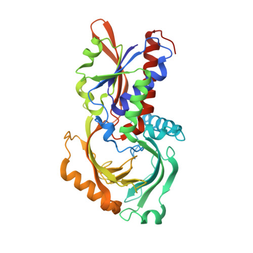
    3WGT

  • PubMed Abstract: 

    The deracemization of racemic amines to yield enantioenriched amines using S-stereoselective amine oxidases (AOx) has recently been attracting attention. However, R-stereoselective AOx that are suitable for deracemization have not yet been identified. An R-stereoselective AOx was now evolved from porcine kidney D-amino acid oxidase (pkDAO) and subsequently use for the deracemization of racemic amines. The engineered pkDAO, which was obtained by directed evolution, displayed a markedly changed substrate specificity towards R amines. The mutant enzyme exhibited a high preference towards the substrate α-methylbenzylamine and was used to synthesize the S amine through deracemization. The findings of this study indicate that further investigations on the structure-activity relationship of AOx are warranted and also provide a new method for biotransformations in organic synthesis.


  • Organizational Affiliation

    Biotechnology Research Center and Department of Biotechnology, Toyama Prefectural University, 5180 Kurokawa, Imizu, Toyama 939-0398 (Japan); Asano Active Enzyme Molecule Project, ERATO, JST, 5180 Kurokawa, Imizu, Toyama 939-0398 (Japan).


Macromolecules
Find similar proteins by:  (by identity cutoff)  |  3D Structure
Entity ID: 1
MoleculeChains Sequence LengthOrganismDetailsImage
D-amino-acid oxidase
A, B
347Sus scrofaMutation(s): 2 
Gene Names: DAO
EC: 1.4.3.3
UniProt
Find proteins for P00371 (Sus scrofa)
Explore P00371 
Go to UniProtKB:  P00371
Entity Groups  
Sequence Clusters30% Identity50% Identity70% Identity90% Identity95% Identity100% Identity
UniProt GroupP00371
Sequence Annotations
Expand
  • Reference Sequence
Small Molecules
Ligands 3 Unique
IDChains Name / Formula / InChI Key2D Diagram3D Interactions
FAD
Query on FAD

Download Ideal Coordinates CCD File 
C [auth A],
G [auth B]
FLAVIN-ADENINE DINUCLEOTIDE
C27 H33 N9 O15 P2
VWWQXMAJTJZDQX-UYBVJOGSSA-N
QSC
Query on QSC

Download Ideal Coordinates CCD File 
D [auth A],
H [auth B]
(1R)-1-phenylethanamine
C8 H11 N
RQEUFEKYXDPUSK-SSDOTTSWSA-N
SO4
Query on SO4

Download Ideal Coordinates CCD File 
E [auth A],
F [auth A],
I [auth B],
J [auth B]
SULFATE ION
O4 S
QAOWNCQODCNURD-UHFFFAOYSA-L
Experimental Data & Validation

Experimental Data

  • Method: X-RAY DIFFRACTION
  • Resolution: 1.88 Å
  • R-Value Free: 0.230 
  • R-Value Work: 0.188 
  • R-Value Observed: 0.191 
  • Space Group: P 21 21 21
Unit Cell:
Length ( Å )Angle ( ˚ )
a = 68.617α = 90
b = 91.607β = 90
c = 110.319γ = 90
Software Package:
Software NamePurpose
HKL-2000data collection
MOLREPphasing
REFMACrefinement
HKL-2000data reduction
HKL-2000data scaling

Structure Validation

View Full Validation Report


Ligand Structure Quality Assessment 


Entry History 

Deposition Data

Revision History  (Full details and data files)

  • Version 1.0: 2014-04-02
    Type: Initial release
  • Version 1.1: 2022-08-24
    Changes: Database references, Derived calculations
  • Version 1.2: 2023-11-08
    Changes: Data collection, Refinement description