3W9W

Crystal structure of DING protein


Experimental Data Snapshot

  • Method: X-RAY DIFFRACTION
  • Resolution: 1.35 Å
  • R-Value Free: 0.170 
  • R-Value Work: 0.148 
  • R-Value Observed: 0.149 

wwPDB Validation   3D Report Full Report


This is version 1.1 of the entry. See complete history


Literature

Crystal structure analysis, overexpression and refolding behaviour of a DING protein with single mutation.

Gai, Z.Q.Nakamura, A.Tanaka, Y.Hirano, N.Tanaka, I.Yao, M.

(2013) J Synchrotron Radiat 20: 854-858

  • DOI: https://doi.org/10.1107/S0909049513020694
  • Primary Citation of Related Structures:  
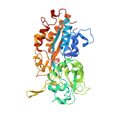
    3W9V, 3W9W

  • PubMed Abstract: 

    After crystallization of a certain protein-RNA complex, well diffracting crystals were obtained. However, the asymmetric unit of the crystal was too small to locate any components. Mass spectrometry and X-ray crystal structure analysis showed that it was a member of the DING protein family (HPBP). Surprisingly, the structure of HPBP reported previously was also determined accidentally as a contaminant, suggesting that HPBP has a strong tendency to crystallize. Furthermore, DING proteins were reported to relate in disease. These observations suggest that DING has potential for application in a wide range of research fields. To enable further analyses, a system for preparation of HPBP was constructed. As HPBP was expressed in insoluble form in Escherichia coli, it was unfolded chemically and refolded. Finally, a very high yield preparation method was constructed, in which 43 mg of HPBP was obtained from 1 L of culture. Furthermore, to evaluate the validity of refolding, its crystal structure was determined at 1.03 Å resolution. The determined structure was identical to the native structure, in which two disulfide bonds were recovered correctly and a phosphate ion was captured. Based on these results, it was concluded that the refolded HPBP recovers its structure correctly.


  • Organizational Affiliation

    Faculty of Advanced Life Sciences, Hokkaido University, Sapporo 060-0810, Japan.


Macromolecules
Find similar proteins by:  (by identity cutoff)  |  3D Structure
Entity ID: 1
MoleculeChains Sequence LengthOrganismDetailsImage
DING protein
A, B
376unidentified prokaryotic organismMutation(s): 1 
UniProt
Find proteins for P85173 (Unknown prokaryotic organism)
Explore P85173 
Go to UniProtKB:  P85173
Entity Groups  
Sequence Clusters30% Identity50% Identity70% Identity90% Identity95% Identity100% Identity
UniProt GroupP85173
Sequence Annotations
Expand
  • Reference Sequence
Small Molecules
Ligands 2 Unique
IDChains Name / Formula / InChI Key2D Diagram3D Interactions
PO4
Query on PO4

Download Ideal Coordinates CCD File 
C [auth A],
H [auth B]
PHOSPHATE ION
O4 P
NBIIXXVUZAFLBC-UHFFFAOYSA-K
GOL
Query on GOL

Download Ideal Coordinates CCD File 
D [auth A]
E [auth A]
F [auth A]
G [auth A]
I [auth B]
D [auth A],
E [auth A],
F [auth A],
G [auth A],
I [auth B],
J [auth B],
K [auth B],
L [auth B]
GLYCEROL
C3 H8 O3
PEDCQBHIVMGVHV-UHFFFAOYSA-N
Experimental Data & Validation

Experimental Data

  • Method: X-RAY DIFFRACTION
  • Resolution: 1.35 Å
  • R-Value Free: 0.170 
  • R-Value Work: 0.148 
  • R-Value Observed: 0.149 
  • Space Group: C 1 2 1
Unit Cell:
Length ( Å )Angle ( ˚ )
a = 94.27α = 90
b = 87.14β = 90.82
c = 88.6γ = 90
Software Package:
Software NamePurpose
XSCALEdata scaling
PHENIXrefinement
PDB_EXTRACTdata extraction
XDSdata reduction
XDSdata scaling

Structure Validation

View Full Validation Report


Entry History 

Deposition Data

Revision History  (Full details and data files)

  • Version 1.0: 2013-10-30
    Type: Initial release
  • Version 1.1: 2023-11-08
    Changes: Data collection, Database references, Derived calculations, Refinement description