3W5R

Crystal structure of complexes of vitamin D receptor ligand binding domain with lithocholic acid derivatives


Experimental Data Snapshot

  • Method: X-RAY DIFFRACTION
  • Resolution: 2.20 Å
  • R-Value Free: 0.276 
  • R-Value Work: 0.220 
  • R-Value Observed: 0.226 

wwPDB Validation   3D Report Full Report


Ligand Structure Quality Assessment 


This is version 1.3 of the entry. See complete history


Literature

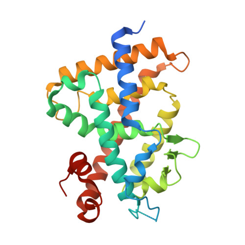
Crystal structures of complexes of vitamin D receptor ligand-binding domain with lithocholic acid derivatives.

Masuno, H.Ikura, T.Morizono, D.Orita, I.Yamada, S.Shimizu, M.Ito, N.

(2013) J Lipid Res 54: 2206-2213

  • DOI: https://doi.org/10.1194/jlr.M038307
  • Primary Citation of Related Structures:  
    3W5P, 3W5Q, 3W5R, 3W5T

  • PubMed Abstract: 

    The secondary bile acid lithocholic acid (LCA) and its derivatives act as selective modulators of the vitamin D receptor (VDR), although their structures fundamentally differ from that of the natural hormone 1α,25-dihydroxyvitamin D3 [1,25(OH)2D3)]. Here, we have determined the crystal structures of the ligand-binding domain of rat VDR (VDR-LBD) in ternary complexes with a synthetic partial peptide of the coactivator MED1 (mediator of RNA polymerase II transcription subunit 1) and four ligands, LCA, 3-keto LCA, LCA acetate, and LCA propionate, with the goal of elucidating their agonistic mechanism. LCA and its derivatives bind to the same ligand-binding pocket (LBP) of VDR-LBD that 1,25(OH)2D3 binds to, but in the opposite orientation; their A-ring is positioned at the top of the LBP, whereas their acyclic tail is located at the bottom of the LBP. However, most of the hydrophobic and hydrophilic interactions observed in the complex with 1,25(OH)2D3 are reproduced in the complexes with LCA and its derivatives. Additional interactions between VDR-LBD and the C-3 substituents of the A-ring are also observed in the complexes with LCA and its derivatives. These may result in the observed difference in the potency among the LCA-type ligands.


  • Organizational Affiliation

    Institute of Biomaterials and Bioengineering, Tokyo Medical and Dental University, Chiyoda-ku, Tokyo 101-0062, Japan.


Macromolecules
Find similar proteins by:  (by identity cutoff)  |  3D Structure
Entity ID: 1
MoleculeChains Sequence LengthOrganismDetailsImage
Vitamin D3 receptor271Rattus norvegicusMutation(s): 0 
Gene Names: VdrNr1i1
UniProt
Find proteins for P13053 (Rattus norvegicus)
Explore P13053 
Go to UniProtKB:  P13053
Entity Groups  
Sequence Clusters30% Identity50% Identity70% Identity90% Identity95% Identity100% Identity
UniProt GroupP13053
Sequence Annotations
Expand
  • Reference Sequence

Find similar proteins by:  Sequence   |   3D Structure  

Entity ID: 2
MoleculeChains Sequence LengthOrganismDetailsImage
Mediator of RNA polymerase II transcription subunit 1B [auth C]13Homo sapiensMutation(s): 0 
UniProt & NIH Common Fund Data Resources
Find proteins for Q15648 (Homo sapiens)
Explore Q15648 
Go to UniProtKB:  Q15648
PHAROS:  Q15648
GTEx:  ENSG00000125686 
Entity Groups  
Sequence Clusters30% Identity50% Identity70% Identity90% Identity95% Identity100% Identity
UniProt GroupQ15648
Sequence Annotations
Expand
  • Reference Sequence
Small Molecules
Ligands 1 Unique
IDChains Name / Formula / InChI Key2D Diagram3D Interactions
LOA
Query on LOA

Download Ideal Coordinates CCD File 
C [auth A](3beta,5beta,9beta)-3-(acetyloxy)cholan-24-oic acid
C26 H42 O4
FCQRLHQHKFKTQE-HCTDMSSWSA-N
Experimental Data & Validation

Experimental Data

  • Method: X-RAY DIFFRACTION
  • Resolution: 2.20 Å
  • R-Value Free: 0.276 
  • R-Value Work: 0.220 
  • R-Value Observed: 0.226 
  • Space Group: C 1 2 1
Unit Cell:
Length ( Å )Angle ( ˚ )
a = 154.433α = 90
b = 42.026β = 96.25
c = 41.514γ = 90
Software Package:
Software NamePurpose
SERGUIdata collection
CNSrefinement
HKL-2000data reduction
HKL-2000data scaling
CNSphasing

Structure Validation

View Full Validation Report


Ligand Structure Quality Assessment 


Entry History 

Deposition Data

Revision History  (Full details and data files)

  • Version 1.0: 2013-06-26
    Type: Initial release
  • Version 1.1: 2014-01-29
    Changes: Database references
  • Version 1.2: 2017-11-22
    Changes: Refinement description
  • Version 1.3: 2023-11-08
    Changes: Data collection, Database references, Derived calculations, Refinement description