3VRB

Mitochondrial rhodoquinol-fumarate reductase from the parasitic nematode Ascaris suum with the specific inhibitor flutolanil and substrate fumarate


Experimental Data Snapshot

  • Method: X-RAY DIFFRACTION
  • Resolution: 2.91 Å
  • R-Value Free: 0.272 
  • R-Value Work: 0.204 
  • R-Value Observed: 0.207 

wwPDB Validation   3D Report Full Report


Ligand Structure Quality Assessment 


This is version 2.0 of the entry. See complete history


Literature

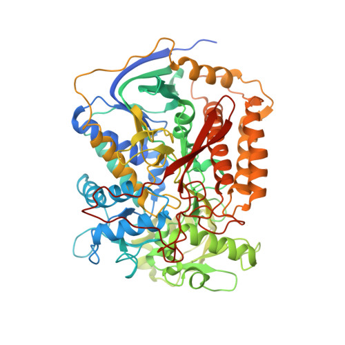
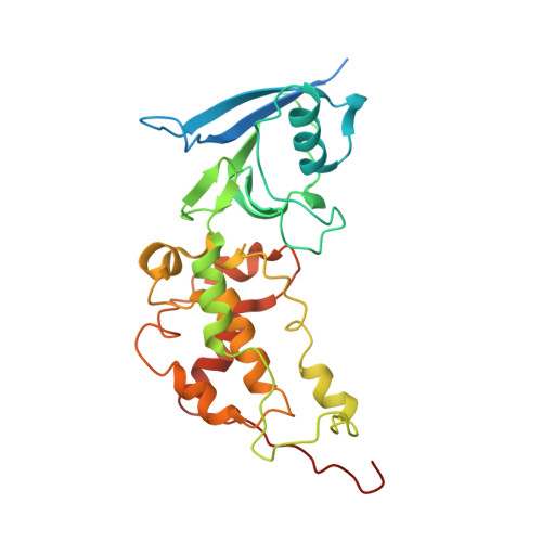
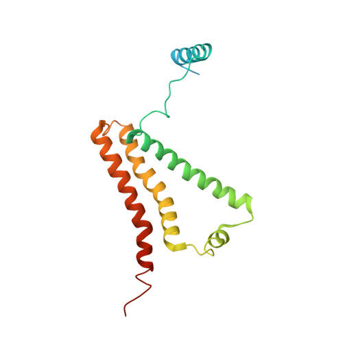
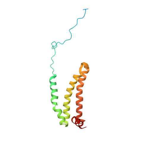
Crystal structure of mitochondrial quinol-fumarate reductase from the parasitic nematode Ascaris suum

Shimizu, H.Osanai, A.Sakamoto, K.Inaoka, D.K.Shiba, T.Harada, S.Kita, K.

(2012) J Biochem 151: 589-592

  • DOI: https://doi.org/10.1093/jb/mvs051
  • Primary Citation of Related Structures:  
    3VR8, 3VRB

  • PubMed Abstract: 

    In the anaerobic respiratory chain of the parasitic nematode Ascaris suum, complex II couples the reduction of fumarate to the oxidation of rhodoquinol, a reverse reaction catalyzed by mammalian complex II. In this study, the first structure of anaerobic complex II of mitochondria was determined. The structure, composed of four subunits and five co-factors, is similar to that of aerobic complex II, except for an extra peptide found in the smallest anchor subunit of the A. suum enzyme. We discuss herein the structure-function relationship of the enzyme and the critical role of the low redox potential of rhodoquinol in the fumarate reduction of A. suum complex II.


  • Organizational Affiliation

    Department of Biomedical Chemistry, Graduate School of Medicine, University of Tokyo, Tokyo, Japan.


Macromolecules
Find similar proteins by:  (by identity cutoff)  |  3D Structure
Entity ID: 1
MoleculeChains Sequence LengthOrganismDetailsImage
Flavoprotein subunit of complex II
A, E
645Ascaris suumMutation(s): 0 
Membrane Entity: Yes 
UniProt
Find proteins for Q33862 (Ascaris suum)
Explore Q33862 
Go to UniProtKB:  Q33862
Entity Groups  
Sequence Clusters30% Identity50% Identity70% Identity90% Identity95% Identity100% Identity
UniProt GroupQ33862
Sequence Annotations
Expand
  • Reference Sequence
Find similar proteins by:  (by identity cutoff)  |  3D Structure
Entity ID: 2
MoleculeChains Sequence LengthOrganismDetailsImage
Iron-sulfur subunit of succinate dehydrogenase
B, F
282Ascaris suumMutation(s): 0 
Membrane Entity: Yes 
UniProt
Find proteins for O44074 (Ascaris suum)
Explore O44074 
Go to UniProtKB:  O44074
Entity Groups  
Sequence Clusters30% Identity50% Identity70% Identity90% Identity95% Identity100% Identity
UniProt GroupO44074
Sequence Annotations
Expand
  • Reference Sequence
Find similar proteins by:  (by identity cutoff)  |  3D Structure
Entity ID: 3
MoleculeChains Sequence LengthOrganismDetailsImage
Cytochrome b-large subunit
C, G
188Ascaris suumMutation(s): 0 
Membrane Entity: Yes 
UniProt
Find proteins for P92506 (Ascaris suum)
Explore P92506 
Go to UniProtKB:  P92506
Entity Groups  
Sequence Clusters30% Identity50% Identity70% Identity90% Identity95% Identity100% Identity
UniProt GroupP92506
Sequence Annotations
Expand
  • Reference Sequence
Find similar proteins by:  (by identity cutoff)  |  3D Structure
Entity ID: 4
MoleculeChains Sequence LengthOrganismDetailsImage
Succinate dehydrogenase [ubiquinone] cytochrome b small subunit, mitochondrial
D, H
156Ascaris suumMutation(s): 0 
Membrane Entity: Yes 
UniProt
Find proteins for P92507 (Ascaris suum)
Explore P92507 
Go to UniProtKB:  P92507
Entity Groups  
Sequence Clusters30% Identity50% Identity70% Identity90% Identity95% Identity100% Identity
UniProt GroupP92507
Sequence Annotations
Expand
  • Reference Sequence
Small Molecules
Ligands 8 Unique
IDChains Name / Formula / InChI Key2D Diagram3D Interactions
FAD
Query on FAD

Download Ideal Coordinates CCD File 
J [auth A],
R [auth E]
FLAVIN-ADENINE DINUCLEOTIDE
C27 H33 N9 O15 P2
VWWQXMAJTJZDQX-UYBVJOGSSA-N
EPH
Query on EPH

Download Ideal Coordinates CCD File 
P [auth D],
X [auth H]
L-ALPHA-PHOSPHATIDYL-BETA-OLEOYL-GAMMA-PALMITOYL-PHOSPHATIDYLETHANOLAMINE
C39 H68 N O8 P
MABRTXOVHMDVAT-AAEGOEIASA-N
HEM
Query on HEM

Download Ideal Coordinates CCD File 
N [auth C],
W [auth G]
PROTOPORPHYRIN IX CONTAINING FE
C34 H32 Fe N4 O4
KABFMIBPWCXCRK-RGGAHWMASA-L
SF4
Query on SF4

Download Ideal Coordinates CCD File 
L [auth B],
T [auth F]
IRON/SULFUR CLUSTER
Fe4 S4
LJBDFODJNLIPKO-UHFFFAOYSA-N
FTN
Query on FTN

Download Ideal Coordinates CCD File 
O [auth C],
V [auth F]
N-[3-(1-methylethoxy)phenyl]-2-(trifluoromethyl)benzamide
C17 H16 F3 N O2
PTCGDEVVHUXTMP-UHFFFAOYSA-N
F3S
Query on F3S

Download Ideal Coordinates CCD File 
M [auth B],
U [auth F]
FE3-S4 CLUSTER
Fe3 S4
FCXHZBQOKRZXKS-UHFFFAOYSA-N
FES
Query on FES

Download Ideal Coordinates CCD File 
K [auth B],
S [auth F]
FE2/S2 (INORGANIC) CLUSTER
Fe2 S2
NIXDOXVAJZFRNF-UHFFFAOYSA-N
FUM
Query on FUM

Download Ideal Coordinates CCD File 
I [auth A],
Q [auth E]
FUMARIC ACID
C4 H4 O4
VZCYOOQTPOCHFL-OWOJBTEDSA-N
Experimental Data & Validation

Experimental Data

  • Method: X-RAY DIFFRACTION
  • Resolution: 2.91 Å
  • R-Value Free: 0.272 
  • R-Value Work: 0.204 
  • R-Value Observed: 0.207 
  • Space Group: P 21 21 21
Unit Cell:
Length ( Å )Angle ( ˚ )
a = 124.306α = 90
b = 131.649β = 90
c = 222.519γ = 90
Software Package:
Software NamePurpose
MOLREPphasing
REFMACrefinement
HKL-2000data reduction
HKL-2000data scaling

Structure Validation

View Full Validation Report


Ligand Structure Quality Assessment 


Entry History 

Deposition Data

Revision History  (Full details and data files)

  • Version 1.0: 2012-07-11
    Type: Initial release
  • Version 1.1: 2012-07-18
    Changes: Non-polymer description
  • Version 1.2: 2012-10-17
    Changes: Atomic model
  • Version 1.3: 2014-12-17
    Changes: Data collection
  • Version 1.4: 2015-06-03
    Changes: Non-polymer description
  • Version 1.5: 2017-11-22
    Changes: Refinement description
  • Version 2.0: 2023-11-15
    Changes: Atomic model, Data collection, Database references, Derived calculations, Structure summary