3VLF

Crystal structure of yeast proteasome interacting protein


Experimental Data Snapshot

  • Method: X-RAY DIFFRACTION
  • Resolution: 3.80 Å
  • R-Value Free: 0.278 
  • R-Value Work: 0.251 
  • R-Value Observed: 0.251 

wwPDB Validation   3D Report Full Report


This is version 1.3 of the entry. See complete history


Literature

Structural basis for specific recognition of Rpt1, an ATPase subunit of the 26S proteasome, by a proteasome-dedicated chaperone Hsm3

Takagi, K.Kim, S.Yukii, H.Ueno, M.Morishita, R.Endo, Y.Kato, K.Tanaka, K.Saeki, Y.Mizushima, T.

(2012) J Biol Chem 287: 12172-12182

  • DOI: https://doi.org/10.1074/jbc.M112.345876
  • Primary Citation of Related Structures:  
    3VLD, 3VLE, 3VLF

  • PubMed Abstract: 

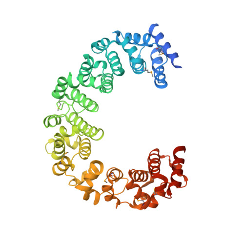
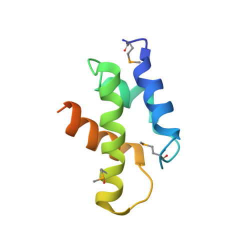
    The 26 S proteasome is a 2.5-MDa molecular machine that degrades ubiquitinated proteins in eukaryotic cells. It consists of a proteolytic core particle and two 19 S regulatory particles (RPs) composed of 6 ATPase (Rpt) and 13 non-ATPase (Rpn) subunits. Multiple proteasome-dedicated chaperones facilitate the assembly of the proteasome, but little is known about the detailed mechanisms. Hsm3, a 19 S RP dedicated chaperone, transiently binds to the C-terminal domain of the Rpt1 subunit and forms a tetrameric complex, Hsm3-Rpt1-Rpt2-Rpn1, during maturation of the ATPase ring of 19 S RP. To elucidate the structural basis of Hsm3 function, we determined the crystal structures of Hsm3 and its complex with the C-terminal domain of the Rpt1 subunit (Rpt1C). Hsm3 has a C-shaped structure that consists of 11 HEAT repeats. The structure of the Hsm3-Rpt1C complex revealed that the interacting surface between Hsm3 and Rpt1 is a hydrophobic core and a complementary charged surface. Mutations in the Hsm3-Rpt1 surface resulted in the assembly defect of the 26 S proteasome. Furthermore, a structural model of the Hsm3-Rpt ring complex and an in vitro binding assay suggest that Hsm3 can bind Rpt2 in addition to Rpt1. Collectively, our results provide the structural basis of the molecular functions of Hsm3 for the RP assembly.


  • Organizational Affiliation

    Picobiology Institute, Department of Life Science, Graduate School of Life Science, University of Hyogo, 3-2-1, Kouto, Kamigori-cho, Ako-gun, Hyogo, 678-1297, Japan.


Macromolecules
Find similar proteins by:  (by identity cutoff)  |  3D Structure
Entity ID: 1
MoleculeChains Sequence LengthOrganismDetailsImage
DNA mismatch repair protein HSM3
A, C
500Saccharomyces cerevisiae S288CMutation(s): 0 
Gene Names: HSM3YBR272CYBR1740
UniProt
Find proteins for P38348 (Saccharomyces cerevisiae (strain ATCC 204508 / S288c))
Explore P38348 
Go to UniProtKB:  P38348
Entity Groups  
Sequence Clusters30% Identity50% Identity70% Identity90% Identity95% Identity100% Identity
UniProt GroupP38348
Sequence Annotations
Expand
  • Reference Sequence
Find similar proteins by:  (by identity cutoff)  |  3D Structure
Entity ID: 2
MoleculeChains Sequence LengthOrganismDetailsImage
26S protease regulatory subunit 7 homolog
B, D
88Saccharomyces cerevisiae S288CMutation(s): 0 
Gene Names: RPT1CIM5YTA3YKL145W
UniProt
Find proteins for P33299 (Saccharomyces cerevisiae (strain ATCC 204508 / S288c))
Explore P33299 
Go to UniProtKB:  P33299
Entity Groups  
Sequence Clusters30% Identity50% Identity70% Identity90% Identity95% Identity100% Identity
UniProt GroupP33299
Sequence Annotations
Expand
  • Reference Sequence
Small Molecules
Modified Residues  1 Unique
IDChains TypeFormula2D DiagramParent
MSE
Query on MSE
A, C
L-PEPTIDE LINKINGC5 H11 N O2 SeMET
Experimental Data & Validation

Experimental Data

  • Method: X-RAY DIFFRACTION
  • Resolution: 3.80 Å
  • R-Value Free: 0.278 
  • R-Value Work: 0.251 
  • R-Value Observed: 0.251 
  • Space Group: P 65 2 2
Unit Cell:
Length ( Å )Angle ( ˚ )
a = 187.282α = 90
b = 187.282β = 90
c = 379.574γ = 120
Software Package:
Software NamePurpose
MLPHAREphasing
CNSrefinement
HKL-2000data reduction
HKL-2000data scaling

Structure Validation

View Full Validation Report


Entry History 

Deposition Data

Revision History  (Full details and data files)

  • Version 1.0: 2012-02-22
    Type: Initial release
  • Version 1.1: 2012-04-18
    Changes: Database references
  • Version 1.2: 2023-11-08
    Changes: Data collection, Database references, Derived calculations, Refinement description
  • Version 1.3: 2023-12-06
    Changes: Data collection