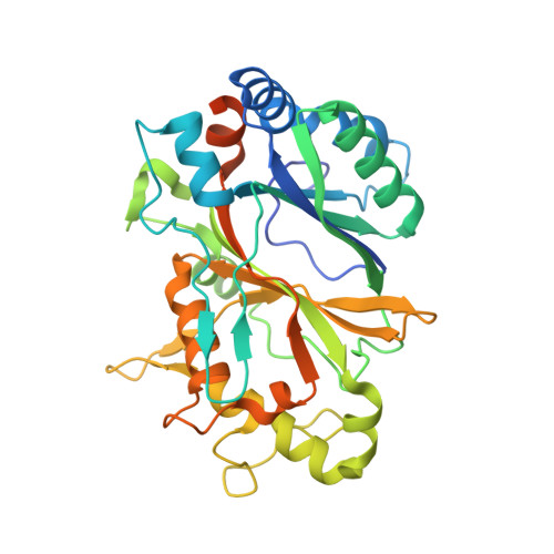
3VED

Rhodococcus jostii RHA1 DypB D153H variant in complex with heme


Experimental Data Snapshot

  • Method: X-RAY DIFFRACTION
  • Resolution: 2.50 Å
  • R-Value Free: 0.218 
  • R-Value Work: 0.184 
  • R-Value Observed: 0.185 

wwPDB Validation   3D Report Full Report


Ligand Structure Quality Assessment 


This is version 1.3 of the entry. See complete history


Literature

Distal heme pocket residues of B-type dye-decolorizing peroxidase: arginine but not aspartate is essential for peroxidase activity.

Singh, R.Grigg, J.C.Armstrong, Z.Murphy, M.E.Eltis, L.D.

(2012) J Biol Chem 287: 10623-10630

  • DOI: https://doi.org/10.1074/jbc.M111.332171
  • Primary Citation of Related Structures:  
    3VEC, 3VED, 3VEE, 3VEF, 3VEG

  • PubMed Abstract: 

    DypB from Rhodococcus jostii RHA1 is a bacterial dye-decolorizing peroxidase (DyP) that oxidizes lignin and Mn(II). Three residues interact with the iron-bound solvent species in ferric DypB: Asn-246 and the conserved Asp-153 and Arg-244. Substitution of either Asp-153 or Asn-246 with alanine minimally affected the second order rate constant for Compound I formation (k(1) ∼ 10(5) M(-1)s(-1)) and the specificity constant (k(cat)/K(m)) for H(2)O(2). Even in the D153A/N246A double variant, these values were reduced less than 30-fold. However, these substitutions dramatically reduced the stability of Compound I (t(1/2) ∼ 0.13 s) as compared with the wild-type enzyme (540 s). By contrast, substitution of Arg-244 with leucine abolished the peroxidase activity, and heme iron of the variant showed a pH-dependent transition from high spin (pH 5) to low spin (pH 8.5). Two variants were designed to mimic the plant peroxidase active site: D153H, which was more than an order of magnitude less reactive with H(2)O(2), and N246H, which had no detectable peroxidase activity. X-ray crystallographic studies revealed that structural changes in the variants are confined to the distal heme environment. The data establish an essential role for Arg-244 in Compound I formation in DypB, possibly through charge stabilization and proton transfer. The principle roles of Asp-153 and Asn-246 appear to be in modulating the subsequent reactivity of Compound I. These results expand the range of residues known to catalyze Compound I formation in heme peroxidases.


  • Organizational Affiliation

    Department of Microbiology and Immunology, Life Sciences Institute, University of British Columbia, Vancouver, British Columbia V6T 1Z3, Canada.


Macromolecules
Find similar proteins by:  (by identity cutoff)  |  3D Structure
Entity ID: 1
MoleculeChains Sequence LengthOrganismDetailsImage
DypB
A, B, C
353Rhodococcus jostii RHA1Mutation(s): 1 
Gene Names: dypBRHA1_ro02407
EC: 1.11.1
UniProt
Find proteins for Q0SE24 (Rhodococcus jostii (strain RHA1))
Explore Q0SE24 
Go to UniProtKB:  Q0SE24
Entity Groups  
Sequence Clusters30% Identity50% Identity70% Identity90% Identity95% Identity100% Identity
UniProt GroupQ0SE24
Sequence Annotations
Expand
  • Reference Sequence
Small Molecules
Ligands 3 Unique
IDChains Name / Formula / InChI Key2D Diagram3D Interactions
HEM
Query on HEM

Download Ideal Coordinates CCD File 
D [auth A],
H [auth B],
K [auth C]
PROTOPORPHYRIN IX CONTAINING FE
C34 H32 Fe N4 O4
KABFMIBPWCXCRK-RGGAHWMASA-L
GOL
Query on GOL

Download Ideal Coordinates CCD File 
F [auth A]GLYCEROL
C3 H8 O3
PEDCQBHIVMGVHV-UHFFFAOYSA-N
CL
Query on CL

Download Ideal Coordinates CCD File 
E [auth A]
G [auth A]
I [auth B]
J [auth B]
L [auth C]
E [auth A],
G [auth A],
I [auth B],
J [auth B],
L [auth C],
M [auth C]
CHLORIDE ION
Cl
VEXZGXHMUGYJMC-UHFFFAOYSA-M
Experimental Data & Validation

Experimental Data

  • Method: X-RAY DIFFRACTION
  • Resolution: 2.50 Å
  • R-Value Free: 0.218 
  • R-Value Work: 0.184 
  • R-Value Observed: 0.185 
  • Space Group: P 32 2 1
Unit Cell:
Length ( Å )Angle ( ˚ )
a = 132.642α = 90
b = 132.642β = 90
c = 159.61γ = 120
Software Package:
Software NamePurpose
SCALEPACKdata scaling
REFMACrefinement
PDB_EXTRACTdata extraction
MxDCdata collection
HKL-2000data reduction
HKL-2000data scaling
REFMACphasing

Structure Validation

View Full Validation Report


Ligand Structure Quality Assessment 


Entry History 

Deposition Data

Revision History  (Full details and data files)

  • Version 1.0: 2012-01-18
    Type: Initial release
  • Version 1.1: 2012-08-29
    Changes: Database references
  • Version 1.2: 2017-11-08
    Changes: Refinement description
  • Version 1.3: 2023-09-13
    Changes: Data collection, Database references, Derived calculations, Refinement description