3VB8

Crystal Structure of Engineered Protein, Northeast Structural Genomics Consortium Target OR43


Experimental Data Snapshot

  • Method: X-RAY DIFFRACTION
  • Resolution: 2.90 Å
  • R-Value Free: 0.290 
  • R-Value Work: 0.244 
  • R-Value Observed: 0.244 

wwPDB Validation   3D Report Full Report


This is version 1.3 of the entry. See complete history


Literature

Computational design of a protein-based enzyme inhibitor.

Procko, E.Hedman, R.Hamilton, K.Seetharaman, J.Fleishman, S.J.Su, M.Aramini, J.Kornhaber, G.Hunt, J.F.Tong, L.Montelione, G.T.Baker, D.

(2013) J Mol Biol 425: 3563-3575

  • DOI: https://doi.org/10.1016/j.jmb.2013.06.035
  • Primary Citation of Related Structures:  
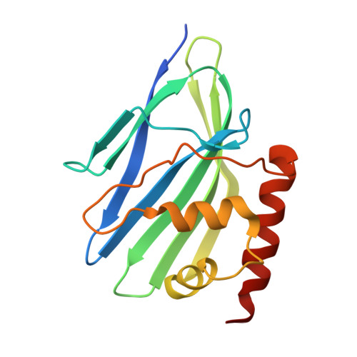
    3VB8

  • PubMed Abstract: 

    While there has been considerable progress in designing protein-protein interactions, the design of proteins that bind polar surfaces is an unmet challenge. We describe the computational design of a protein that binds the acidic active site of hen egg lysozyme and inhibits the enzyme. The design process starts with two polar amino acids that fit deep into the enzyme active site, identifies a protein scaffold that supports these residues and is complementary in shape to the lysozyme active-site region, and finally optimizes the surrounding contact surface for high-affinity binding. Following affinity maturation, a protein designed using this method bound lysozyme with low nanomolar affinity, and a combination of NMR studies, crystallography, and knockout mutagenesis confirmed the designed binding surface and orientation. Saturation mutagenesis with selection and deep sequencing demonstrated that specific designed interactions extending well beyond the centrally grafted polar residues are critical for high-affinity binding.


  • Organizational Affiliation

    Department of Biochemistry and Howard Hughes Medical Institute, University of Washington, Seattle, WA 98195, USA. procko@u.washington.edu


Macromolecules
Find similar proteins by:  (by identity cutoff)  |  3D Structure
Entity ID: 1
MoleculeChains Sequence LengthOrganismDetailsImage
Engineered protein
A, B
161synthetic constructMutation(s): 0 
Entity Groups  
Sequence Clusters30% Identity50% Identity70% Identity90% Identity95% Identity100% Identity
Sequence Annotations
Expand
  • Reference Sequence
Small Molecules
Ligands 1 Unique
IDChains Name / Formula / InChI Key2D Diagram3D Interactions
SO4
Query on SO4

Download Ideal Coordinates CCD File 
C [auth A]SULFATE ION
O4 S
QAOWNCQODCNURD-UHFFFAOYSA-L
Experimental Data & Validation

Experimental Data

  • Method: X-RAY DIFFRACTION
  • Resolution: 2.90 Å
  • R-Value Free: 0.290 
  • R-Value Work: 0.244 
  • R-Value Observed: 0.244 
  • Space Group: P 43 21 2
Unit Cell:
Length ( Å )Angle ( ˚ )
a = 77.553α = 90
b = 77.553β = 90
c = 115.741γ = 90
Software Package:
Software NamePurpose
ADSCdata collection
MOLREPphasing
CNSrefinement
HKL-2000data reduction
HKL-2000data scaling

Structure Validation

View Full Validation Report


Entry History 

Revision History  (Full details and data files)

  • Version 1.0: 2012-06-06
    Type: Initial release
  • Version 1.1: 2013-07-24
    Changes: Database references
  • Version 1.2: 2013-09-11
    Changes: Database references
  • Version 1.3: 2023-09-13
    Changes: Data collection, Database references, Derived calculations, Refinement description