3V57

Crystal Structure of the B-phycoerythrin from the red algae Porphyridium Cruentum at pH8


Experimental Data Snapshot

  • Method: X-RAY DIFFRACTION
  • Resolution: 1.70 Å
  • R-Value Free: 0.215 
  • R-Value Work: 0.177 
  • R-Value Observed: 0.179 

wwPDB Validation   3D Report Full Report


Ligand Structure Quality Assessment 


This is version 1.1 of the entry. See complete history


Literature

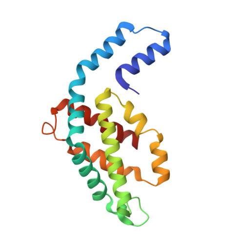
pH-dependent structural conformations of B-phycoerythrin from Porphyridium cruentum

Camara-Artigas, A.Bacarizo, J.Andujar-Sanchez, M.Ortiz-Salmeron, E.Mesa-Valle, C.Cuadri, C.Martin-Garcia, J.M.Martinez-Rodriguez, S.Mazzuca-Sobczuk, T.Ibanez, M.J.Allen, J.P.

(2012) FEBS J 279: 3680-3691

  • DOI: https://doi.org/10.1111/j.1742-4658.2012.08730.x
  • Primary Citation of Related Structures:  
    3V57, 3V58

  • PubMed Abstract: 

    B-phycoerythrin from the red alga Porphyridium cruentum was crystallized using the technique of capillary counter-diffusion. Crystals belonging to the space group R3 with almost identical unit cell constants and diffracting to 1.85 and 1.70 Å were obtained at pH values of 5 and 8, respectively. The most important difference between structures is the presence of the residue His88α in two different conformations at pH 8. This residue is placed next to the chromophore phycoerythrobilin PEB82α and the new conformation results in the relocation of the hydrogen-bond network and hydration around PEB82α, which probably contributes to the observed pH dependence of the optical spectrum associated with this chromophore. Comparison with the structures of B-phycoerythrin from other red algae shows differences in the conformation of the A-ring of the chromophore PEB139α. This conformational difference in B-phycoerythrin from P. cruentum enables the formation of several hydrogen bonds that connect PEB139α with the chromophore PEB158β at the (αβ)(3) hexamer association interface. The possible influence of these structural differences on the optical spectrum and the ability of the protein to perform energy transfer are discussed, with the two pH-dependent conformations of His88α and PEB82α being proposed as representing critical structural features that are correlated with the pH dependence of the optical spectrum and transient optical states during energy transfer.


  • Organizational Affiliation

    Department of Physical Chemistry, Biochemistry and Inorganic Chemistry, Agrifood Campus of International Excellence (CeiA3), University of Almería, Spain.


Macromolecules
Find similar proteins by:  (by identity cutoff)  |  3D Structure
Entity ID: 1
MoleculeChains Sequence LengthOrganismDetailsImage
Phycoerythrin alpha subunit
A, C
164Porphyridium purpureumMutation(s): 0 
UniProt
Find proteins for P11392 (Porphyridium purpureum)
Explore P11392 
Go to UniProtKB:  P11392
Entity Groups  
Sequence Clusters30% Identity50% Identity70% Identity90% Identity95% Identity100% Identity
UniProt GroupP11392
Sequence Annotations
Expand
  • Reference Sequence
Find similar proteins by:  (by identity cutoff)  |  3D Structure
Entity ID: 2
MoleculeChains Sequence LengthOrganismDetailsImage
Phycoerythrin beta subunit
B, D
177Porphyridium purpureumMutation(s): 0 
UniProt
Find proteins for P11393 (Porphyridium purpureum)
Explore P11393 
Go to UniProtKB:  P11393
Entity Groups  
Sequence Clusters30% Identity50% Identity70% Identity90% Identity95% Identity100% Identity
UniProt GroupP11393
Sequence Annotations
Expand
  • Reference Sequence
Small Molecules
Ligands 2 Unique
IDChains Name / Formula / InChI Key2D Diagram3D Interactions
PEB
Query on PEB

Download Ideal Coordinates CCD File 
E [auth A]
F [auth A]
I [auth B]
J [auth B]
K [auth B]
E [auth A],
F [auth A],
I [auth B],
J [auth B],
K [auth B],
M [auth C],
N [auth C],
O [auth D],
P [auth D],
Q [auth D]
PHYCOERYTHROBILIN
C33 H40 N4 O6
NKCBCVIFPXGHAV-WAVSMFBNSA-N
SO4
Query on SO4

Download Ideal Coordinates CCD File 
G [auth A],
H [auth A],
L [auth B],
R [auth D],
S [auth D]
SULFATE ION
O4 S
QAOWNCQODCNURD-UHFFFAOYSA-L
Modified Residues  1 Unique
IDChains TypeFormula2D DiagramParent
MEN
Query on MEN
B, D
L-PEPTIDE LINKINGC5 H10 N2 O3ASN
Experimental Data & Validation

Experimental Data

  • Method: X-RAY DIFFRACTION
  • Resolution: 1.70 Å
  • R-Value Free: 0.215 
  • R-Value Work: 0.177 
  • R-Value Observed: 0.179 
  • Space Group: H 3
Unit Cell:
Length ( Å )Angle ( ˚ )
a = 187.11α = 90
b = 187.11β = 90
c = 59.23γ = 120
Software Package:
Software NamePurpose
MOSFLMdata reduction
SCALAdata scaling
MOLREPphasing
REFMACrefinement
PDB_EXTRACTdata extraction
ADSCdata collection

Structure Validation

View Full Validation Report


Ligand Structure Quality Assessment 


Entry History 

Deposition Data

Revision History  (Full details and data files)

  • Version 1.0: 2012-10-03
    Type: Initial release
  • Version 1.1: 2023-09-13
    Changes: Data collection, Database references, Derived calculations, Refinement description