3V3X

Nitroxide Spin Labels in Protein GB1: N8/K28 Double Mutant


Experimental Data Snapshot

  • Method: X-RAY DIFFRACTION
  • Resolution: 2.00 Å
  • R-Value Free: 0.288 
  • R-Value Work: 0.228 
  • R-Value Observed: 0.231 

wwPDB Validation   3D Report Full Report


Ligand Structure Quality Assessment 


This is version 1.2 of the entry. See complete history


Literature

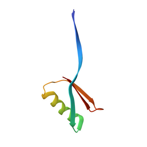
High-resolution structure of a protein spin-label in a solvent-exposed beta-sheet and comparison with DEER spectroscopy.

Cunningham, T.F.McGoff, M.S.Sengupta, I.Jaroniec, C.P.Horne, W.S.Saxena, S.

(2012) Biochemistry 51: 6350-6359

  • DOI: https://doi.org/10.1021/bi300328w
  • Primary Citation of Related Structures:  
    3V3X

  • PubMed Abstract: 

    X-ray crystallography has been a useful tool in the development of site-directed spin labeling by resolving rotamers of the nitroxide spin-label side chain in a variety of α-helical environments. In this work, the crystal structure of a doubly spin-labeled N8C/K28C mutant of the B1 immunoglobulin-binding domain of protein G (GB1) was solved. The double mutant formed a domain-swapped dimer under crystallization conditions. Two rotameric states of the spin-label were resolved at the solvent-exposed α-helical site, at residue 28; these are in good agreement with rotamers previously reported for helical structures. The second site, at residue 8 on an interior β-strand, shows the presence of three distinct solvent-exposed side-chain rotamers. One of these rotamers is rarely observed within crystal structures of R1 sites and suggests that the H(α) and S(δ) hydrogen bond that is common to α-helical sites is absent at this interior β-strand residue. Variable temperature continuous wave (CW) experiments of the β-strand site showed two distinct components that were correlated to the rotameric states observed in crystallography. Interestingly, the CW data at room temperature could be fit without the use of an order parameter, which is consistent with the lack of the H(α) and S(δ) interaction. Additionally, double electron electron resonance (DEER) spectroscopy was performed on the GB1 double mutant in its monomeric form and yielded a most probable interspin distance of 25 ± 1 Å. In order to evaluate the accuracy of the measured DEER distance, the rotamers observed in the crystal structure of the domain-swapped GB1 dimer were modeled into a high-resolution structure of the wild type monomeric GB1. The distances generated in the resulting GB1 structural models match the most probable DEER distance within ~2 Å. The results are interesting as they indicate by direct experimental measurement that the rotameric states of R1 found in this crystal provide a very close match to the most probable distance measured by DEER.


  • Organizational Affiliation

    Department of Chemistry, University of Pittsburgh, 219 Parkman Avenue, Pittsburgh, PA 15260, USA.


Macromolecules
Find similar proteins by:  (by identity cutoff)  |  3D Structure
Entity ID: 1
MoleculeChains Sequence LengthOrganismDetailsImage
Immunoglobulin G-binding protein G
A, B, C, D
56Streptococcus sp. 'group GMutation(s): 2 
Gene Names: spg
UniProt
Find proteins for P19909 (Streptococcus sp. group G)
Explore P19909 
Go to UniProtKB:  P19909
Entity Groups  
Sequence Clusters30% Identity50% Identity70% Identity90% Identity95% Identity100% Identity
UniProt GroupP19909
Sequence Annotations
Expand
  • Reference Sequence
Small Molecules
Ligands 4 Unique
IDChains Name / Formula / InChI Key2D Diagram3D Interactions
2PE
Query on 2PE

Download Ideal Coordinates CCD File 
F [auth A]NONAETHYLENE GLYCOL
C18 H38 O10
YZUUTMGDONTGTN-UHFFFAOYSA-N
MTN
Query on MTN

Download Ideal Coordinates CCD File 
E [auth A]
G [auth B]
H [auth B]
K [auth C]
L [auth C]
E [auth A],
G [auth B],
H [auth B],
K [auth C],
L [auth C],
M [auth D],
N [auth D]
S-[(1-oxyl-2,2,5,5-tetramethyl-2,5-dihydro-1H-pyrrol-3-yl)methyl] methanesulfonothioate
C10 H18 N O3 S2
MXZPGYFBZHBAQM-UHFFFAOYSA-N
GOL
Query on GOL

Download Ideal Coordinates CCD File 
I [auth B]GLYCEROL
C3 H8 O3
PEDCQBHIVMGVHV-UHFFFAOYSA-N
ACT
Query on ACT

Download Ideal Coordinates CCD File 
J [auth B],
O [auth D]
ACETATE ION
C2 H3 O2
QTBSBXVTEAMEQO-UHFFFAOYSA-M
Experimental Data & Validation

Experimental Data

  • Method: X-RAY DIFFRACTION
  • Resolution: 2.00 Å
  • R-Value Free: 0.288 
  • R-Value Work: 0.228 
  • R-Value Observed: 0.231 
  • Space Group: C 1 2 1
Unit Cell:
Length ( Å )Angle ( ˚ )
a = 105.248α = 90
b = 35.747β = 126.37
c = 86.989γ = 90
Software Package:
Software NamePurpose
StructureStudiodata collection
PHASERphasing
REFMACrefinement
d*TREKdata reduction
d*TREKdata scaling

Structure Validation

View Full Validation Report


Ligand Structure Quality Assessment 


Entry History 

Deposition Data

Revision History  (Full details and data files)

  • Version 1.0: 2012-08-29
    Type: Initial release
  • Version 1.1: 2013-06-19
    Changes: Database references
  • Version 1.2: 2023-09-13
    Changes: Advisory, Data collection, Database references, Derived calculations, Refinement description