Metal-mediated affinity and orientation specificity in a computationally designed protein homodimer.
Der, B.S., Machius, M., Miley, M.J., Mills, J.L., Szyperski, T., Kuhlman, B.(2012) J Am Chem Soc 134: 375-385
- PubMed: 22092237
- DOI: https://doi.org/10.1021/ja208015j
- Primary Citation of Related Structures:
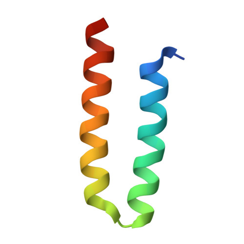
3V1A, 3V1B, 3V1C, 3V1D, 3V1E, 3V1F - PubMed Abstract:
Computationally designing protein-protein interactions with high affinity and desired orientation is a challenging task. Incorporating metal-binding sites at the target interface may be one approach for increasing affinity and specifying the binding mode, thereby improving robustness of designed interactions for use as tools in basic research as well as in applications from biotechnology to medicine. Here we describe a Rosetta-based approach for the rational design of a protein monomer to form a zinc-mediated, symmetric homodimer. Our metal interface design, named MID1 (NESG target ID OR37), forms a tight dimer in the presence of zinc (MID1-zinc) with a dissociation constant <30 nM. Without zinc the dissociation constant is 4 μM. The crystal structure of MID1-zinc shows good overall agreement with the computational model, but only three out of four designed histidines coordinate zinc. However, a histidine-to-glutamate point mutation resulted in four-coordination of zinc, and the resulting metal binding site and dimer orientation closely matches the computational model (Cα rmsd = 1.4 Å).
Organizational Affiliation:
Department of Biochemistry and Biophysics, University of North Carolina, Chapel Hill, North Carolina 27599-7260, USA.