3UV0

Crystal structure of the drosophila MU2 FHA domain


Experimental Data Snapshot

  • Method: X-RAY DIFFRACTION
  • Resolution: 1.90 Å
  • R-Value Free: 0.227 
  • R-Value Work: 0.178 
  • R-Value Observed: 0.180 

wwPDB Validation   3D Report Full Report


This is version 1.3 of the entry. See complete history


Literature

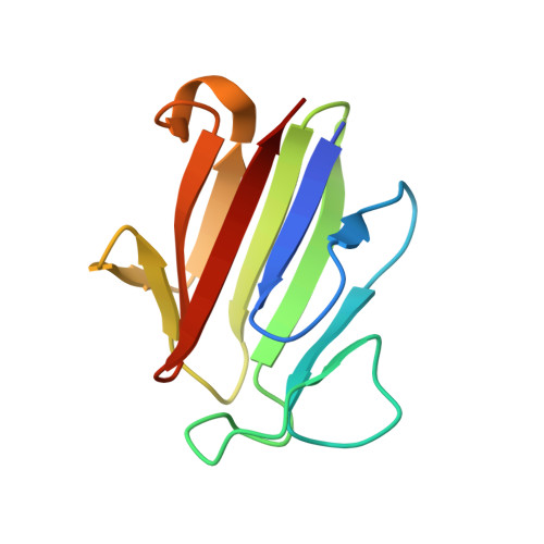
Dimerization, but not phosphothreonine binding, is conserved between the forkhead-associated domains of Drosophila MU2 and human MDC1

Luo, S.Ye, K.

(2012) FEBS Lett 586: 344-349

  • DOI: https://doi.org/10.1016/j.febslet.2012.01.023
  • Primary Citation of Related Structures:  
    3UV0

  • PubMed Abstract: 

    Mutator 2 (MU2) in Drosophila melanogaster has been proposed to be the ortholog of human MDC1, a key mediator in DNA damage response. The forkhead-associated (FHA) domain of MDC1 is a dimerization module regulated by trans binding to phosphothreonine 4 from another molecule. Here we present the crystal structure of the MU2 FHA domain at 1.9Å resolution, revealing its evolutionarily conserved role in dimerization. As compared to the MDC1 FHA domain, the MU2 FHA domain dimerizes using a different and more stable interface and contains a degenerate phosphothreonine-binding pocket. Our results suggest that the MU2 dimerization is constitutive and lacks phosphorylation-mediated regulation.


  • Organizational Affiliation

    College of Biological Sciences, China Agricultural University, Beijing, China.


Macromolecules
Find similar proteins by:  (by identity cutoff)  |  3D Structure
Entity ID: 1
MoleculeChains Sequence LengthOrganismDetailsImage
Mutator 2, isoform B
A, B
102Drosophila melanogasterMutation(s): 0 
Gene Names: mu2Dmel_CG1960
UniProt
Find proteins for Q9W061 (Drosophila melanogaster)
Explore Q9W061 
Go to UniProtKB:  Q9W061
Entity Groups  
Sequence Clusters30% Identity50% Identity70% Identity90% Identity95% Identity100% Identity
UniProt GroupQ9W061
Sequence Annotations
Expand
  • Reference Sequence
Experimental Data & Validation

Experimental Data

  • Method: X-RAY DIFFRACTION
  • Resolution: 1.90 Å
  • R-Value Free: 0.227 
  • R-Value Work: 0.178 
  • R-Value Observed: 0.180 
  • Space Group: P 65
Unit Cell:
Length ( Å )Angle ( ˚ )
a = 99.86α = 90
b = 99.86β = 90
c = 38.31γ = 120
Software Package:
Software NamePurpose
REFMACrefinement
PDB_EXTRACTdata extraction
HKL-2000data reduction
SCALEPACKdata scaling
SHARPphasing

Structure Validation

View Full Validation Report


Entry History 

Deposition Data

  • Released Date: 2012-01-25 
  • Deposition Author(s): Luo, S., Ye, K.

Revision History  (Full details and data files)

  • Version 1.0: 2012-01-25
    Type: Initial release
  • Version 1.1: 2012-02-22
    Changes: Database references
  • Version 1.2: 2014-03-12
    Changes: Database references
  • Version 1.3: 2024-03-20
    Changes: Data collection, Database references