3UTF

Crystal structure of Aspergillus fumigatus UDP galactopyranose mutase in reduced state


Experimental Data Snapshot

  • Method: X-RAY DIFFRACTION
  • Resolution: 2.25 Å
  • R-Value Free: 0.215 
  • R-Value Work: 0.190 
  • R-Value Observed: 0.191 

wwPDB Validation   3D Report Full Report


Ligand Structure Quality Assessment 


This is version 1.3 of the entry. See complete history


Literature

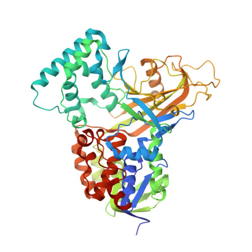
Crystal Structures and Small-angle X-ray Scattering Analysis of UDP-galactopyranose Mutase from the Pathogenic Fungus Aspergillus fumigatus.

Dhatwalia, R.Singh, H.Oppenheimer, M.Karr, D.B.Nix, J.C.Sobrado, P.Tanner, J.J.

(2012) J Biol Chem 287: 9041-9051

  • DOI: https://doi.org/10.1074/jbc.M111.327536
  • Primary Citation of Related Structures:  
    3UTE, 3UTF, 3UTG, 3UTH

  • PubMed Abstract: 

    UDP-galactopyranose mutase (UGM) is a flavoenzyme that catalyzes the conversion of UDP-galactopyranose to UDP-galactofuranose, which is a central reaction in galactofuranose biosynthesis. Galactofuranose has never been found in humans but is an essential building block of the cell wall and extracellular matrix of many bacteria, fungi, and protozoa. The importance of UGM for the viability of many pathogens and its absence in humans make UGM a potential drug target. Here we report the first crystal structures and small-angle x-ray scattering data for UGM from the fungus Aspergillus fumigatus, the causative agent of aspergillosis. The structures reveal that Aspergillus UGM has several extra secondary and tertiary structural elements that are not found in bacterial UGMs yet are important for substrate recognition and oligomerization. Small-angle x-ray scattering data show that Aspergillus UGM forms a tetramer in solution, which is unprecedented for UGMs. The binding of UDP or the substrate induces profound conformational changes in the enzyme. Two loops on opposite sides of the active site move toward each other by over 10 Å to cover the substrate and create a closed active site. The degree of substrate-induced conformational change exceeds that of bacterial UGMs and is a direct consequence of the unique quaternary structure of Aspergillus UGM. Galactopyranose binds at the re face of the FAD isoalloxazine with the anomeric carbon atom poised for nucleophilic attack by the FAD N5 atom. The structural data provide new insight into substrate recognition and the catalytic mechanism and thus will aid inhibitor design.


  • Organizational Affiliation

    Department of Chemistry, University of Missouri, Columbia, Missouri 65211, USA.


Macromolecules
Find similar proteins by:  (by identity cutoff)  |  3D Structure
Entity ID: 1
MoleculeChains Sequence LengthOrganismDetailsImage
UDP-galactopyranose mutase
A, B, C, D
513Aspergillus fumigatusMutation(s): 2 
Gene Names: glfglfA
EC: 5.4.99.9
UniProt
Find proteins for Q4W1X2 (Aspergillus fumigatus)
Explore Q4W1X2 
Go to UniProtKB:  Q4W1X2
Entity Groups  
Sequence Clusters30% Identity50% Identity70% Identity90% Identity95% Identity100% Identity
UniProt GroupQ4W1X2
Sequence Annotations
Expand
  • Reference Sequence
Small Molecules
Ligands 2 Unique
IDChains Name / Formula / InChI Key2D Diagram3D Interactions
FDA
Query on FDA

Download Ideal Coordinates CCD File 
E [auth A],
L [auth B],
R [auth C],
Z [auth D]
DIHYDROFLAVINE-ADENINE DINUCLEOTIDE
C27 H35 N9 O15 P2
YPZRHBJKEMOYQH-UYBVJOGSSA-N
SO4
Query on SO4

Download Ideal Coordinates CCD File 
AA [auth D]
BA [auth D]
CA [auth D]
DA [auth D]
EA [auth D]
AA [auth D],
BA [auth D],
CA [auth D],
DA [auth D],
EA [auth D],
F [auth A],
G [auth A],
H [auth A],
I [auth A],
J [auth A],
K [auth A],
M [auth B],
N [auth B],
O [auth B],
P [auth B],
Q [auth B],
S [auth C],
T [auth C],
U [auth C],
V [auth C],
W [auth C],
X [auth C],
Y [auth C]
SULFATE ION
O4 S
QAOWNCQODCNURD-UHFFFAOYSA-L
Experimental Data & Validation

Experimental Data

  • Method: X-RAY DIFFRACTION
  • Resolution: 2.25 Å
  • R-Value Free: 0.215 
  • R-Value Work: 0.190 
  • R-Value Observed: 0.191 
  • Space Group: P 65 2 2
Unit Cell:
Length ( Å )Angle ( ˚ )
a = 217.965α = 90
b = 217.965β = 90
c = 322.805γ = 120
Software Package:
Software NamePurpose
SCALAdata scaling
PHENIXrefinement
PDB_EXTRACTdata extraction
ADSCdata collection
XDSdata reduction
PHENIXphasing

Structure Validation

View Full Validation Report


Ligand Structure Quality Assessment 


Entry History 

Deposition Data

Revision History  (Full details and data files)

  • Version 1.0: 2012-02-08
    Type: Initial release
  • Version 1.1: 2012-02-15
    Changes: Database references
  • Version 1.2: 2012-04-04
    Changes: Database references
  • Version 1.3: 2023-09-13
    Changes: Data collection, Database references, Derived calculations, Refinement description