3UMM

Formylglycinamide ribonucleotide amidotransferase from Salmonella typhimurium: Role of the ATP complexation and glutaminase domain in catalytic coupling


Experimental Data Snapshot

  • Method: X-RAY DIFFRACTION
  • Resolution: 3.20 Å
  • R-Value Free: 0.276 
  • R-Value Work: 0.196 

wwPDB Validation   3D Report Full Report


Ligand Structure Quality Assessment 


This is version 1.1 of the entry. See complete history


Literature

Formylglycinamide ribonucleotide amidotransferase from Salmonella typhimurium: role of ATP complexation and the glutaminase domain in catalytic coupling

Tanwar, A.S.Morar, M.Panjikar, S.Anand, R.

(2012) Acta Crystallogr D Biol Crystallogr 68: 627-636

  • DOI: https://doi.org/10.1107/S0907444912006543
  • Primary Citation of Related Structures:  
    3UGJ, 3UJN, 3UMM

  • PubMed Abstract: 

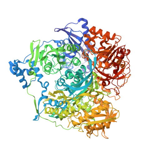
    Formylglycinamide ribonucleotide (FGAR) amidotransferase (FGAR-AT) takes part in purine biosynthesis and is a multidomain enzyme with multiple spatially separated active sites. FGAR-AT contains a glutaminase domain that is responsible for the generation of ammonia from glutamine. Ammonia is then transferred via a channel to a second active site located in the synthetase domain and utilized to convert FGAR to formylglycinamidine ribonucleotide (FGAM) in an adenosine triphosphate (ATP) dependent reaction. In some ammonia-channelling enzymes ligand binding triggers interdomain signalling between the two diverse active centres and also assists in formation of the ammonia channel. Previously, the structure of FGAR-AT from Salmonella typhimurium containing a glutamyl thioester intermediate covalently bound in the glutaminase active site was determined. In this work, the roles played by various ligands of FGAR-AT in inducing catalytic coupling are investigated. Structures of FGAR-AT from S. typhimurium were determined in two different states: the unliganded form and the binary complex with an ATP analogue in the presence of the glutamyl thioester intermediate. The structures were compared in order to decipher the roles of these two states in interdomain communication. Using a process of elimination, the results indicated that binding of FGAR is most likely to be the major mechanism by which catalytic coupling occurs. This is because conformational changes do not occur either upon formation of the glutamyl thioester intermediate or upon subsequent ATP complexation. A model of the FGAR-bound form of the enzyme suggested that the loop in the synthetase domain may be responsible for initiating catalytic coupling via its interaction with the N-terminal domain.


  • Organizational Affiliation

    Department of Chemistry, Indian Institute of Technology, IIT-Bombay, Mumbai 400 076, India.


Macromolecules
Find similar proteins by:  (by identity cutoff)  |  3D Structure
Entity ID: 1
MoleculeChains Sequence LengthOrganismDetailsImage
Phosphoribosylformylglycinamidine synthase1,303Salmonella enterica subsp. enterica serovar TyphimuriumMutation(s): 0 
EC: 6.3.5.3
UniProt
Find proteins for P74881 (Salmonella typhimurium (strain LT2 / SGSC1412 / ATCC 700720))
Explore P74881 
Go to UniProtKB:  P74881
Entity Groups  
Sequence Clusters30% Identity50% Identity70% Identity90% Identity95% Identity100% Identity
UniProt GroupP74881
Sequence Annotations
Expand
  • Reference Sequence
Small Molecules
Ligands 4 Unique
IDChains Name / Formula / InChI Key2D Diagram3D Interactions
ANP
Query on ANP

Download Ideal Coordinates CCD File 
B [auth A]PHOSPHOAMINOPHOSPHONIC ACID-ADENYLATE ESTER
C10 H17 N6 O12 P3
PVKSNHVPLWYQGJ-KQYNXXCUSA-N
ADP
Query on ADP

Download Ideal Coordinates CCD File 
C [auth A]ADENOSINE-5'-DIPHOSPHATE
C10 H15 N5 O10 P2
XTWYTFMLZFPYCI-KQYNXXCUSA-N
SO4
Query on SO4

Download Ideal Coordinates CCD File 
G [auth A],
H [auth A]
SULFATE ION
O4 S
QAOWNCQODCNURD-UHFFFAOYSA-L
MG
Query on MG

Download Ideal Coordinates CCD File 
D [auth A],
E [auth A],
F [auth A]
MAGNESIUM ION
Mg
JLVVSXFLKOJNIY-UHFFFAOYSA-N
Modified Residues  1 Unique
IDChains TypeFormula2D DiagramParent
CYG
Query on CYG
A
L-PEPTIDE LINKINGC8 H14 N2 O5 SCYS
Experimental Data & Validation

Experimental Data

  • Method: X-RAY DIFFRACTION
  • Resolution: 3.20 Å
  • R-Value Free: 0.276 
  • R-Value Work: 0.196 
  • Space Group: P 65
Unit Cell:
Length ( Å )Angle ( ˚ )
a = 148.68α = 90
b = 148.68β = 90
c = 142.05γ = 120
Software Package:
Software NamePurpose
MOSFLMdata reduction
SCALAdata scaling
CNSrefinement
PDB_EXTRACTdata extraction
MAR345dtbdata collection
CNSphasing

Structure Validation

View Full Validation Report


Ligand Structure Quality Assessment 


Entry History 

Deposition Data

Revision History  (Full details and data files)

  • Version 1.0: 2012-06-06
    Type: Initial release
  • Version 1.1: 2013-09-11
    Changes: Database references