3UMD

Structure of pB intermediate of Photoactive yellow protein (PYP) at pH 4.


Experimental Data Snapshot

  • Method: X-RAY DIFFRACTION
  • Resolution: 1.80 Å
  • R-Value Free: 0.238 
  • R-Value Work: 0.196 

wwPDB Validation   3D Report Full Report


Ligand Structure Quality Assessment 


This is version 1.2 of the entry. See complete history


Literature

pH Dependence of the Photoactive Yellow Protein Photocycle Investigated by Time-Resolved Crystallography.

Tripathi, S.Srajer, V.Purwar, N.Henning, R.Schmidt, M.

(2012) Biophys J 102: 325-332

  • DOI: https://doi.org/10.1016/j.bpj.2011.11.4021
  • Primary Citation of Related Structures:  
    3UMD, 3UME

  • PubMed Abstract: 

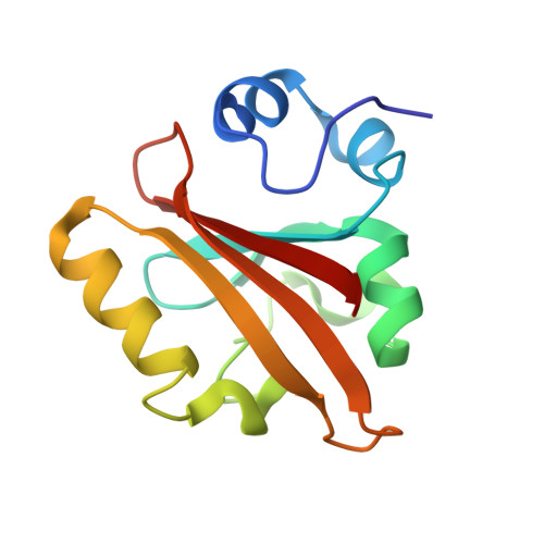
    Visualizing the three-dimensional structures of a protein during its biological activity is key to understanding its mechanism. In general, protein structure and function are pH-dependent. Changing the pH provides new insights into the mechanisms that are involved in protein activity. Photoactive yellow protein (PYP) is a signaling protein that serves as an ideal model for time-dependent studies on light-activated proteins. Its photocycle is studied extensively under different pH conditions. However, the structures of the intermediates remain unknown until time-resolved crystallography is employed. With the newest beamline developments, a comprehensive time series of Laue data can now be collected from a single protein crystal. This allows us to vary the pH. Here we present the first structure, to our knowledge, of a short-lived protein-inhibitor complex formed in the pB state of the PYP photocycle at pH 4. A water molecule that is transiently stabilized in the chromophore active site prevents the relaxation of the chromophore back to the trans configuration. As a result, the dark-state recovery is slowed down dramatically. At pH 9, PYP stops cycling through the pB state altogether. The electrostatic environment in the chromophore-binding site is the likely reason for this altered kinetics at different pH values.


  • Organizational Affiliation

    University of Wisconsin-Milwaukee, Milwaukee, Wisconsin, USA.


Macromolecules
Find similar proteins by:  (by identity cutoff)  |  3D Structure
Entity ID: 1
MoleculeChains Sequence LengthOrganismDetailsImage
Photoactive yellow protein125Halorhodospira halophilaMutation(s): 0 
UniProt
Find proteins for P16113 (Halorhodospira halophila)
Explore P16113 
Go to UniProtKB:  P16113
Entity Groups  
Sequence Clusters30% Identity50% Identity70% Identity90% Identity95% Identity100% Identity
UniProt GroupP16113
Sequence Annotations
Expand
  • Reference Sequence
Small Molecules
Ligands 1 Unique
IDChains Name / Formula / InChI Key2D Diagram3D Interactions
HC4
Query on HC4

Download Ideal Coordinates CCD File 
B [auth A]4'-HYDROXYCINNAMIC ACID
C9 H8 O3
NGSWKAQJJWESNS-ZZXKWVIFSA-N
Experimental Data & Validation

Experimental Data

  • Method: X-RAY DIFFRACTION
  • Resolution: 1.80 Å
  • R-Value Free: 0.238 
  • R-Value Work: 0.196 
  • Space Group: P 63
Unit Cell:
Length ( Å )Angle ( ˚ )
a = 66.9α = 90
b = 66.9β = 90
c = 40.8γ = 120
Software Package:
Software NamePurpose
XFITdata reduction
CNSrefinement
Precognitiondata reduction
Epinormdata reduction

Structure Validation

View Full Validation Report


Ligand Structure Quality Assessment 


Entry History 

Deposition Data

Revision History  (Full details and data files)

  • Version 1.0: 2012-04-11
    Type: Initial release
  • Version 1.1: 2017-11-08
    Changes: Refinement description
  • Version 1.2: 2023-09-13
    Changes: Data collection, Database references, Derived calculations, Refinement description