Evidence in Support of Lysine 77 and Histidine 96 as Acid-Base Catalytic Residues in Saccharopine Dehydrogenase from Saccharomyces cerevisiae.
Kumar, V.P., Thomas, L.M., Bobyk, K.D., Andi, B., Cook, P.F., West, A.H.(2012) Biochemistry 51: 857-866
- PubMed: 22243403
- DOI: https://doi.org/10.1021/bi201808u
- Primary Citation of Related Structures:
3UGK, 3UH1, 3UHA - PubMed Abstract:
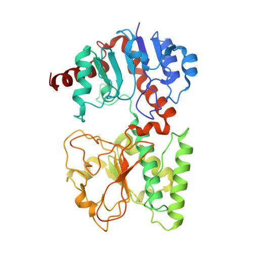
Saccharopine dehydrogenase (SDH) catalyzes the final reaction in the α-aminoadipate pathway, the conversion of l-saccharopine to l-lysine (Lys) and α-ketoglutarate (α-kg) using NAD⁺ as an oxidant. The enzyme utilizes a general acid-base mechanism to conduct its reaction with a base proposed to accept a proton from the secondary amine of saccharopine in the oxidation step and a group proposed to activate water to hydrolyze the resulting imine. Crystal structures of an open apo form and a closed form of the enzyme with saccharopine and NADH bound have been determined at 2.0 and 2.2 Å resolution, respectively. In the ternary complex, a significant movement of domain I relative to domain II that closes the active site cleft between the two domains and brings H96 and K77 into the proximity of the substrate binding site is observed. The hydride transfer distance is 3.6 Å, and the side chains of H96 and K77 are properly positioned to act as acid-base catalysts. Preparation of the K77M and H96Q single-mutant and K77M/H96Q double-mutant enzymes provides data consistent with their role as the general acid-base catalysts in the SDH reaction. The side chain of K77 initially accepts a proton from the ε-amine of the substrate Lys and eventually donates it to the imino nitrogen as it is reduced to a secondary amine in the hydride transfer step, and H96 protonates the carbonyl oxygen as the carbinolamine is formed. The K77M, H976Q, and K77M/H96Q mutant enzymes give 145-, 28-, and 700-fold decreases in V/E(t) and >10³-fold increases in V₂/K(Lys)E(t) and V₂/K(α-kg)E(t) (the double mutation gives >10⁵-fold decreases in the second-order rate constants). In addition, the K77M mutant enzyme exhibits a primary deuterium kinetic isotope effect of 2.0 and an inverse solvent deuterium isotope effect of 0.77 on V₂/K(Lys). A value of 2.0 was also observed for (D)(V₂/K(Lys))(D₂O) when the primary deuterium kinetic isotope effect was repeated in D₂O, consistent with a rate-limiting hydride transfer step. A viscosity effect of 0.8 was observed on V₂/K(Lys), indicating the solvent deuterium isotope effect resulted from stabilization of an enzyme form prior to hydride transfer. A small normal solvent isotope effect is observed on V, which decreases slightly when repeated with NADD, consistent with a contribution from product release to rate limitation. In addition, V₂/K(Lys)E(t) is pH-independent, which is consistent with the loss of an acid-base catalyst and perturbation of the pK(a) of the second catalytic group to a higher pH, likely a result of a change in the overall charge of the active site. The primary deuterium kinetic isotope effect for H96Q, measured in H₂O or D₂O, is within error equal to 1. A solvent deuterium isotope effect of 2.4 is observed with NADH or NADD as the dinucleotide substrate. Data suggest rate-limiting imine formation, consistent with the proposed role of H96 in protonating the leaving hydroxyl as the imine is formed. The pH-rate profile for V₂/K(Lys)E(t) exhibits the pK(a) for K77, perturbed to a value of ∼9, which must be unprotonated to accept a proton from the ε-amine of the substrate Lys so that it can act as a nucleophile. Overall, data are consistent with a role for K77 acting as the base that accepts a proton from the ε-amine of the substrate lysine prior to nucleophilic attack on the α-oxo group of α-ketoglutarate, and finally donating a proton to the imine nitrogen as it is reduced to give saccharopine. In addition, data indicate a role for H96 acting as a general acid-base catalyst in the formation of the imine between the ε-amine of lysine and the α-oxo group of α-ketoglutarate.
Organizational Affiliation:
Department of Chemistry and Biochemistry, University of Oklahoma, Norman, Oklahoma 73019, United States.