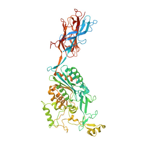
3TXA

Structural Analysis of Adhesive Tip pilin, GBS104 from Group B Streptococcus agalactiae


Experimental Data Snapshot

  • Method: X-RAY DIFFRACTION
  • Resolution: 2.62 Å
  • R-Value Free: 0.256 
  • R-Value Work: 0.206 
  • R-Value Observed: 0.208 

wwPDB Validation   3D Report Full Report


This is version 1.4 of the entry. See complete history


Literature

Structure of Streptococcus agalactiae tip pilin GBS104: a model for GBS pili assembly and host interactions.

Krishnan, V.Dwivedi, P.Kim, B.J.Samal, A.Macon, K.Ma, X.Mishra, A.Doran, K.S.Ton-That, H.Narayana, S.V.

(2013) Acta Crystallogr D Biol Crystallogr 69: 1073-1089

  • DOI: https://doi.org/10.1107/S0907444913004642
  • Primary Citation of Related Structures:  
    3TVY, 3TW0, 3TXA

  • PubMed Abstract: 

    The crystal structure of a 75 kDa central fragment of GBS104, a tip pilin from the 2063V/R strain of Streptococcus agalactiae (group B streptococcus; GBS), is reported. In addition, a homology model of the remaining two domains of GBS104 was built and a model of full-length GBS104 was generated by combining the homology model (the N1 and N4 domains) and the crystal structure of the 75 kDa fragment (the N2 and N3 domains). This rod-shaped GBS104 model is constructed of three IgG-like domains (the N1, N2 and N4 domains) and one vWFA-like domain (the N3 domain). The N1 and N2 domains of GBS104 are assembled with distinct and remote segments contributed by the N- and C-termini. The metal-binding site in the N3 domain of GBS104 is in the closed/low-affinity conformation. Interestingly, this domain hosts two long arms that project away from the metal-binding site. Using site-directed mutagenesis, two cysteine residues that lock the N3 domain of GBS104 into the open/high-affinity conformation were introduced. Both wild-type and disulfide-locked recombinant proteins were tested for binding to extracellular matrix proteins such as collagen, fibronectin, fibrinogen and laminin, and an increase in fibronectin binding affinity was identified for the disulfide-locked N3 domain, suggesting that induced conformational changes may play a possible role in receptor binding.


  • Organizational Affiliation

    UNESCO Regional Centre for Biotechnology (RCB), Gurgaon 122 016, Haryana, India.


Macromolecules
Find similar proteins by:  (by identity cutoff)  |  3D Structure
Entity ID: 1
MoleculeChains Sequence LengthOrganismDetailsImage
Cell wall surface anchor family protein682Streptococcus agalactiae serogroup VMutation(s): 0 
Gene Names: SAG0649
UniProt
Find proteins for Q8E0S5 (Streptococcus agalactiae serotype V (strain ATCC BAA-611 / 2603 V/R))
Explore Q8E0S5 
Go to UniProtKB:  Q8E0S5
Entity Groups  
Sequence Clusters30% Identity50% Identity70% Identity90% Identity95% Identity100% Identity
UniProt GroupQ8E0S5
Sequence Annotations
Expand
  • Reference Sequence
Small Molecules
Ligands 3 Unique
IDChains Name / Formula / InChI Key2D Diagram3D Interactions
CD
Query on CD

Download Ideal Coordinates CCD File 
C [auth A]
D [auth A]
E [auth A]
F [auth A]
G [auth A]
C [auth A],
D [auth A],
E [auth A],
F [auth A],
G [auth A],
H [auth A],
I [auth A]
CADMIUM ION
Cd
WLZRMCYVCSSEQC-UHFFFAOYSA-N
MG
Query on MG

Download Ideal Coordinates CCD File 
B [auth A]MAGNESIUM ION
Mg
JLVVSXFLKOJNIY-UHFFFAOYSA-N
LI
Query on LI

Download Ideal Coordinates CCD File 
J [auth A]LITHIUM ION
Li
HBBGRARXTFLTSG-UHFFFAOYSA-N
Experimental Data & Validation

Experimental Data

  • Method: X-RAY DIFFRACTION
  • Resolution: 2.62 Å
  • R-Value Free: 0.256 
  • R-Value Work: 0.206 
  • R-Value Observed: 0.208 
  • Space Group: P 31 2 1
Unit Cell:
Length ( Å )Angle ( ˚ )
a = 124.585α = 90
b = 124.585β = 90
c = 117.839γ = 120
Software Package:
Software NamePurpose
ADSCdata collection
PHASERphasing
REFMACrefinement
HKL-2000data reduction
HKL-2000data scaling

Structure Validation

View Full Validation Report


Entry History 

Deposition Data

Revision History  (Full details and data files)

  • Version 1.0: 2013-03-27
    Type: Initial release
  • Version 1.1: 2013-05-29
    Changes: Database references
  • Version 1.2: 2013-06-19
    Changes: Database references
  • Version 1.3: 2021-08-25
    Changes: Advisory, Database references, Derived calculations
  • Version 1.4: 2023-09-13
    Changes: Data collection, Refinement description