3TQ0

Crystal structure of Leishmania major dihydroorotate dehydrogenase in complex with fumarate


Experimental Data Snapshot

  • Method: X-RAY DIFFRACTION
  • Resolution: 1.90 Å
  • R-Value Free: 0.182 
  • R-Value Work: 0.165 
  • R-Value Observed: 0.165 

wwPDB Validation   3D Report Full Report


Ligand Structure Quality Assessment 


This is version 2.0 of the entry. See complete history


Literature

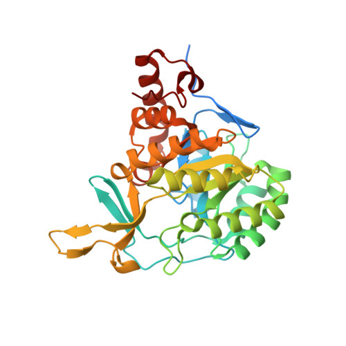
Crystal structure of dihydroorotate dehydrogenase from Leishmania major.

Cordeiro, A.T.Feliciano, P.R.Pinheiro, M.P.Nonato, M.C.

(2012) Biochimie 94: 1739-1748

  • DOI: https://doi.org/10.1016/j.biochi.2012.04.003
  • Primary Citation of Related Structures:  
    3TQ0

  • PubMed Abstract: 

    Dihydroorotate dehydrogenase (DHODH) is the fourth enzyme in the de novo pyrimidine biosynthetic pathway and has been exploited as the target for therapy against proliferative and parasitic diseases. In this study, we report the crystal structures of DHODH from Leishmania major, the species of Leishmania associated with zoonotic cutaneous leishmaniasis, in its apo form and in complex with orotate and fumarate molecules. Both orotate and fumarate were found to bind to the same active site and exploit similar interactions, consistent with a ping-pong mechanism described for class 1A DHODHs. Analysis of LmDHODH structures reveals that rearrangements in the conformation of the catalytic loop have direct influence on the dimeric interface. This is the first structural evidence of a relationship between the dimeric form and the catalytic mechanism. According to our analysis, the high sequence and structural similarity observed among trypanosomatid DHODH suggest that a single strategy of structure-based inhibitor design can be used to validate DHODH as a druggable target against multiple neglected tropical diseases such as Leishmaniasis, Sleeping sickness and Chagas' diseases.


  • Organizational Affiliation

    Laboratório de Cristalografia de Proteínas, Departamento de Física e Química, Faculdade de Ciências Farmacêuticas de Ribeirão Preto, Universidade de São Paulo, Av. Café S/N, Monte Alegre, Ribeirão Preto 14040-903, S.P, Brazil.


Macromolecules
Find similar proteins by:  (by identity cutoff)  |  3D Structure
Entity ID: 1
MoleculeChains Sequence LengthOrganismDetailsImage
Dihydroorotate dehydrogenase
A, B
346Leishmania major strain FriedlinMutation(s): 0 
Gene Names: DHODHlmjf16.0530LMJF_16_0530
EC: 1.3.3.1
UniProt
Find proteins for Q4QEW7 (Leishmania major)
Explore Q4QEW7 
Go to UniProtKB:  Q4QEW7
Entity Groups  
Sequence Clusters30% Identity50% Identity70% Identity90% Identity95% Identity100% Identity
UniProt GroupQ4QEW7
Sequence Annotations
Expand
  • Reference Sequence
Small Molecules
Ligands 5 Unique
IDChains Name / Formula / InChI Key2D Diagram3D Interactions
FMN
Query on FMN

Download Ideal Coordinates CCD File 
C [auth A],
K [auth B]
FLAVIN MONONUCLEOTIDE
C17 H21 N4 O9 P
FVTCRASFADXXNN-SCRDCRAPSA-N
FUM
Query on FUM

Download Ideal Coordinates CCD File 
D [auth A],
L [auth B]
FUMARIC ACID
C4 H4 O4
VZCYOOQTPOCHFL-OWOJBTEDSA-N
SO4
Query on SO4

Download Ideal Coordinates CCD File 
G [auth A],
H [auth A],
I [auth A],
J [auth A]
SULFATE ION
O4 S
QAOWNCQODCNURD-UHFFFAOYSA-L
GOL
Query on GOL

Download Ideal Coordinates CCD File 
E [auth A],
M [auth B]
GLYCEROL
C3 H8 O3
PEDCQBHIVMGVHV-UHFFFAOYSA-N
NI
Query on NI

Download Ideal Coordinates CCD File 
F [auth A]NICKEL (II) ION
Ni
VEQPNABPJHWNSG-UHFFFAOYSA-N
Experimental Data & Validation

Experimental Data

  • Method: X-RAY DIFFRACTION
  • Resolution: 1.90 Å
  • R-Value Free: 0.182 
  • R-Value Work: 0.165 
  • R-Value Observed: 0.165 
  • Space Group: P 61
Unit Cell:
Length ( Å )Angle ( ˚ )
a = 140.833α = 90
b = 140.833β = 90
c = 68.348γ = 120
Software Package:
Software NamePurpose
MAR345dtbdata collection
MOLREPphasing
REFMACrefinement
MOSFLMdata reduction
SCALAdata scaling

Structure Validation

View Full Validation Report


Ligand Structure Quality Assessment 


Entry History 

Deposition Data

Revision History  (Full details and data files)

  • Version 1.0: 2012-05-16
    Type: Initial release
  • Version 1.1: 2012-07-04
    Changes: Database references
  • Version 1.2: 2012-07-25
    Changes: Non-polymer description
  • Version 1.3: 2023-09-13
    Changes: Data collection, Database references, Derived calculations, Refinement description
  • Version 2.0: 2023-11-15
    Changes: Atomic model, Data collection