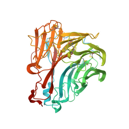
3TIA

Crystal structure of 1957 pandemic H2N2 neuraminidase complexed with laninamivir


Experimental Data Snapshot

  • Method: X-RAY DIFFRACTION
  • Resolution: 1.80 Å
  • R-Value Free: 0.170 
  • R-Value Work: 0.152 
  • R-Value Observed: 0.152 

wwPDB Validation   3D Report Full Report


Ligand Structure Quality Assessment 


This is version 2.1 of the entry. See complete history


Literature

Structural and functional analysis of laninamivir and its octanoate prodrug reveals group specific mechanisms for influenza NA inhibition

Vavricka, C.J.Li, Q.Wu, Y.Qi, J.Wang, M.Liu, Y.Gao, F.Liu, J.Feng, E.He, J.Wang, J.Liu, H.Jiang, H.Gao, G.F.

(2011) PLoS Pathog 7: e1002249-e1002249

  • DOI: https://doi.org/10.1371/journal.ppat.1002249
  • Primary Citation of Related Structures:  
    3TI3, 3TI4, 3TI5, 3TI6, 3TI8, 3TIA, 3TIB, 3TIC

  • PubMed Abstract: 

    The 2009 H1N1 influenza pandemic (pH1N1) led to record sales of neuraminidase (NA) inhibitors, which has contributed significantly to the recent increase in oseltamivir-resistant viruses. Therefore, development and careful evaluation of novel NA inhibitors is of great interest. Recently, a highly potent NA inhibitor, laninamivir, has been approved for use in Japan. Laninamivir is effective using a single inhaled dose via its octanoate prodrug (CS-8958) and has been demonstrated to be effective against oseltamivir-resistant NA in vitro. However, effectiveness of laninamivir octanoate prodrug against oseltamivir-resistant influenza infection in adults has not been demonstrated. NA is classified into 2 groups based upon phylogenetic analysis and it is becoming clear that each group has some distinct structural features. Recently, we found that pH1N1 N1 NA (p09N1) is an atypical group 1 NA with some group 2-like features in its active site (lack of a 150-cavity). Furthermore, it has been reported that certain oseltamivir-resistant substitutions in the NA active site are group 1 specific. In order to comprehensively evaluate the effectiveness of laninamivir, we utilized recombinant N5 (typical group 1), p09N1 (atypical group 1) and N2 from the 1957 pandemic H2N2 (p57N2) (typical group 2) to carry out in vitro inhibition assays. We found that laninamivir and its octanoate prodrug display group specific preferences to different influenza NAs and provide the structural basis of their specific action based upon their novel complex crystal structures. Our results indicate that laninamivir and zanamivir are more effective against group 1 NA with a 150-cavity than group 2 NA with no 150-cavity. Furthermore, we have found that the laninamivir octanoate prodrug has a unique binding mode in p09N1 that is different from that of group 2 p57N2, but with some similarities to NA-oseltamivir binding, which provides additional insight into group specific differences of oseltamivir binding and resistance.


  • Organizational Affiliation

    CAS Key Laboratory of Pathogenic Microbiology and Immunology, Institute of Microbiology, Chinese Academy of Sciences, Beijing, China.


Macromolecules
Find similar proteins by:  (by identity cutoff)  |  3D Structure
Entity ID: 1
MoleculeChains Sequence LengthOrganismDetailsImage
Neuraminidase
A, B, C, D
469Influenza A virus (A/RI/5+/1957(H2N2))Mutation(s): 0 
UniProt
Find proteins for Q194T1 (Influenza A virus)
Explore Q194T1 
Go to UniProtKB:  Q194T1
Entity Groups  
Sequence Clusters30% Identity50% Identity70% Identity90% Identity95% Identity100% Identity
UniProt GroupQ194T1
Sequence Annotations
Expand
  • Reference Sequence
Oligosaccharides

Help

Entity ID: 2
MoleculeChains Length2D Diagram Glycosylation3D Interactions
2-acetamido-2-deoxy-beta-D-glucopyranose-(1-4)-2-acetamido-2-deoxy-beta-D-glucopyranose
E, H, J
2N-Glycosylation
Glycosylation Resources
GlyTouCan:  G42666HT
GlyCosmos:  G42666HT
GlyGen:  G42666HT
Entity ID: 3
MoleculeChains Length2D Diagram Glycosylation3D Interactions
alpha-D-mannopyranose-(1-3)-beta-D-mannopyranose-(1-4)-2-acetamido-2-deoxy-beta-D-glucopyranose-(1-4)-2-acetamido-2-deoxy-beta-D-glucopyranose
F, G, I, K
4N-Glycosylation
Glycosylation Resources
GlyTouCan:  G81315DD
GlyCosmos:  G81315DD
GlyGen:  G81315DD
Small Molecules
Ligands 3 Unique
IDChains Name / Formula / InChI Key2D Diagram3D Interactions
LNV
Query on LNV

Download Ideal Coordinates CCD File 
M [auth A],
P [auth B],
R [auth C],
T [auth D]
5-acetamido-2,6-anhydro-4-carbamimidamido-3,4,5-trideoxy-7-O-methyl-D-glycero-D-galacto-non-2-enonic acid
C13 H22 N4 O7
QNRRHYPPQFELSF-CNYIRLTGSA-N
NAG
Query on NAG

Download Ideal Coordinates CCD File 
O [auth B]2-acetamido-2-deoxy-beta-D-glucopyranose
C8 H15 N O6
OVRNDRQMDRJTHS-FMDGEEDCSA-N
CA
Query on CA

Download Ideal Coordinates CCD File 
L [auth A],
N [auth B],
Q [auth C],
S [auth D]
CALCIUM ION
Ca
BHPQYMZQTOCNFJ-UHFFFAOYSA-N
Binding Affinity Annotations 
IDSourceBinding Affinity
LNV Binding MOAD:  3TIA IC50: 3.12 (nM) from 1 assay(s)
PDBBind:  3TIA IC50: 3.12 (nM) from 1 assay(s)
BindingDB:  3TIA IC50: 3100 (nM) from 1 assay(s)
Experimental Data & Validation

Experimental Data

  • Method: X-RAY DIFFRACTION
  • Resolution: 1.80 Å
  • R-Value Free: 0.170 
  • R-Value Work: 0.152 
  • R-Value Observed: 0.152 
  • Space Group: P 1 21 1
Unit Cell:
Length ( Å )Angle ( ˚ )
a = 89.928α = 90
b = 140.305β = 101.54
c = 89.893γ = 90
Software Package:
Software NamePurpose
ADSCdata collection
PHASERphasing
PHENIXrefinement
HKL-2000data reduction
HKL-2000data scaling

Structure Validation

View Full Validation Report


Ligand Structure Quality Assessment 


Entry History 

Deposition Data

Revision History  (Full details and data files)

  • Version 1.0: 2011-11-16
    Type: Initial release
  • Version 2.0: 2020-07-29
    Type: Remediation
    Reason: Carbohydrate remediation
    Changes: Atomic model, Data collection, Derived calculations, Structure summary
  • Version 2.1: 2023-11-01
    Changes: Data collection, Database references, Refinement description, Structure summary