3TEO

APO Form of carbon disulfide hydrolase (selenomethionine form)


Experimental Data Snapshot

  • Method: X-RAY DIFFRACTION
  • Resolution: 2.40 Å
  • R-Value Free: 0.271 
  • R-Value Work: 0.224 
  • R-Value Observed: 0.226 

wwPDB Validation   3D Report Full Report


This is version 1.3 of the entry. See complete history


Literature

Evolution of a new enzyme for carbon disulphide conversion by an acidothermophilic archaeon.

Smeulders, M.J.Barends, T.R.Pol, A.Scherer, A.Zandvoort, M.H.Udvarhelyi, A.Khadem, A.F.Menzel, A.Hermans, J.Shoeman, R.L.Wessels, H.J.van den Heuvel, L.P.Russ, L.Schlichting, I.Jetten, M.S.Op den Camp, H.J.

(2011) Nature 478: 412-416

  • DOI: https://doi.org/10.1038/nature10464
  • Primary Citation of Related Structures:  
    3TEN, 3TEO

  • PubMed Abstract: 

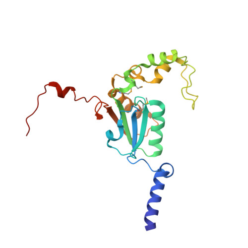
    Extremophilic organisms require specialized enzymes for their exotic metabolisms. Acid-loving thermophilic Archaea that live in the mudpots of volcanic solfataras obtain their energy from reduced sulphur compounds such as hydrogen sulphide (H(2)S) and carbon disulphide (CS(2)). The oxidation of these compounds into sulphuric acid creates the extremely acidic environment that characterizes solfataras. The hyperthermophilic Acidianus strain A1-3, which was isolated from the fumarolic, ancient sauna building at the Solfatara volcano (Naples, Italy), was shown to rapidly convert CS(2) into H(2)S and carbon dioxide (CO(2)), but nothing has been known about the modes of action and the evolution of the enzyme(s) involved. Here we describe the structure, the proposed mechanism and evolution of a CS(2) hydrolase from Acidianus A1-3. The enzyme monomer displays a typical β-carbonic anhydrase fold and active site, yet CO(2) is not one of its substrates. Owing to large carboxy- and amino-terminal arms, an unusual hexadecameric catenane oligomer has evolved. This structure results in the blocking of the entrance to the active site that is found in canonical β-carbonic anhydrases and the formation of a single 15-Å-long, highly hydrophobic tunnel that functions as a specificity filter. The tunnel determines the enzyme's substrate specificity for CS(2), which is hydrophobic. The transposon sequences that surround the gene encoding this CS(2) hydrolase point to horizontal gene transfer as a mechanism for its acquisition during evolution. Our results show how the ancient β-carbonic anhydrase, which is central to global carbon metabolism, was transformed by divergent evolution into a crucial enzyme in CS(2) metabolism.


  • Organizational Affiliation

    Department of Microbiology, Radboud University Nijmegen, Heyendaalseweg 135, 6525 AJ, Nijmegen, The Netherlands.


Macromolecules
Find similar proteins by:  (by identity cutoff)  |  3D Structure
Entity ID: 1
MoleculeChains Sequence LengthOrganismDetailsImage
Carbon disulfide hydrolase
A, B, C, D, E
A, B, C, D, E, F, G, H, I, J, K, L, M, N, O, P
204Acidianus sp. A1-3Mutation(s): 0 
Gene Names: cs2 hydrolase
UniProt
Find proteins for G0WXL9 (Acidianus sp. (strain A1-3))
Explore G0WXL9 
Go to UniProtKB:  G0WXL9
Entity Groups  
Sequence Clusters30% Identity50% Identity70% Identity90% Identity95% Identity100% Identity
UniProt GroupG0WXL9
Sequence Annotations
Expand
  • Reference Sequence
Small Molecules
Ligands 2 Unique
IDChains Name / Formula / InChI Key2D Diagram3D Interactions
PE3
Query on PE3

Download Ideal Coordinates CCD File 
AA [auth G],
MA [auth O]
3,6,9,12,15,18,21,24,27,30,33,36,39-TRIDECAOXAHENTETRACONTANE-1,41-DIOL
C28 H58 O15
ILLKMACMBHTSHP-UHFFFAOYSA-N
CL
Query on CL

Download Ideal Coordinates CCD File 
BA [auth H]
CA [auth I]
DA [auth I]
EA [auth J]
FA [auth K]
BA [auth H],
CA [auth I],
DA [auth I],
EA [auth J],
FA [auth K],
GA [auth L],
HA [auth M],
IA [auth N],
JA [auth N],
KA [auth O],
LA [auth O],
NA [auth P],
Q [auth A],
R [auth B],
S [auth C],
T [auth C],
U [auth C],
V [auth D],
W [auth E],
X [auth E],
Y [auth F],
Z [auth G]
CHLORIDE ION
Cl
VEXZGXHMUGYJMC-UHFFFAOYSA-M
Modified Residues  1 Unique
IDChains TypeFormula2D DiagramParent
MSE
Query on MSE
A, B, C, D, E
A, B, C, D, E, F, G, H, I, J, K, L, M, N, O, P
L-PEPTIDE LINKINGC5 H11 N O2 SeMET
Experimental Data & Validation

Experimental Data

  • Method: X-RAY DIFFRACTION
  • Resolution: 2.40 Å
  • R-Value Free: 0.271 
  • R-Value Work: 0.224 
  • R-Value Observed: 0.226 
  • Space Group: C 1 2 1
Unit Cell:
Length ( Å )Angle ( ˚ )
a = 227.18α = 90
b = 165.51β = 91.17
c = 115.94γ = 90
Software Package:
Software NamePurpose
PHASERphasing
REFMACrefinement
XDSdata reduction
XSCALEdata scaling

Structure Validation

View Full Validation Report


Entry History 

Revision History  (Full details and data files)

  • Version 1.0: 2011-10-19
    Type: Initial release
  • Version 1.1: 2011-10-26
    Changes: Database references
  • Version 1.2: 2012-03-21
    Changes: Database references
  • Version 1.3: 2017-11-08
    Changes: Refinement description, Structure summary