3TD5

Crystal structure of OmpA-like domain from Acinetobacter baumannii in complex with L-Ala-gamma-D-Glu-m-DAP-D-Ala-D-Ala


Experimental Data Snapshot

  • Method: X-RAY DIFFRACTION
  • Resolution: 2.00 Å
  • R-Value Free: 0.257 
  • R-Value Work: 0.220 

wwPDB Validation   3D Report Full Report


This is version 2.0 of the entry. See complete history


Literature

Mechanism of anchoring of OmpA protein to the cell wall peptidoglycan of the gram-negative bacterial outer membrane

Park, J.S.Lee, W.C.Yeo, K.J.Ryu, K.S.Kumarasiri, M.Hesek, D.Lee, M.Mobashery, S.Song, J.H.Kim, S.I.Lee, J.C.Cheong, C.Jeon, Y.H.Kim, H.Y.

(2012) FASEB J 26: 219-228

  • DOI: https://doi.org/10.1096/fj.11-188425
  • Primary Citation of Related Structures:  
    3TD3, 3TD4, 3TD5

  • PubMed Abstract: 

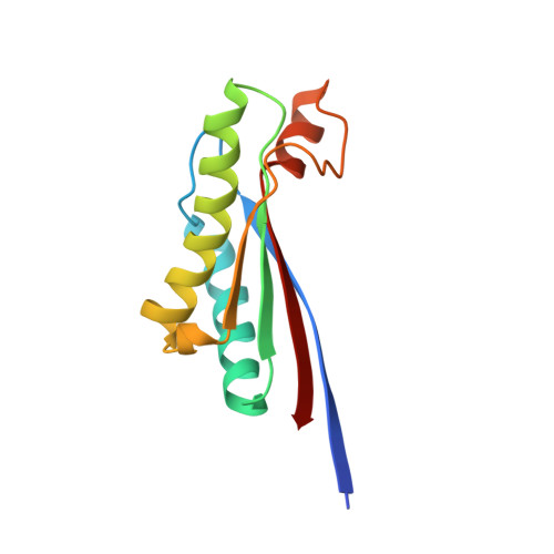
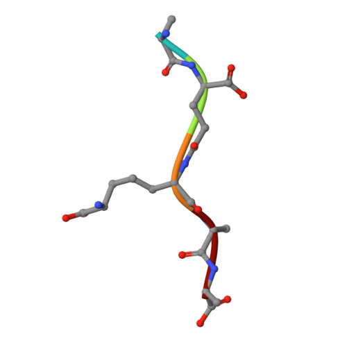
    The outer membrane protein A (OmpA) plays important roles in anchoring of the outer membrane to the bacterial cell wall. The C-terminal periplasmic domain of OmpA (OmpA-like domain) associates with the peptidoglycan (PGN) layer noncovalently. However, there is a paucity of information on the structural aspects of the mechanism of PGN recognition by OmpA-like domains. To elucidate this molecular recognition process, we solved the high-resolution crystal structure of an OmpA-like domain from Acinetobacter baumannii bound to diaminopimelate (DAP), a unique bacterial amino acid from the PGN. The structure clearly illustrates that two absolutely conserved Asp271 and Arg286 residues are the key to the binding to DAP of PGN. Identification of DAP as the central anchoring site of PGN to OmpA is further supported by isothermal titration calorimetry and a pulldown assay with PGN. An NMR-based computational model for complexation between the PGN and OmpA emerged, and this model is validated by determining the crystal structure in complex with a synthetic PGN fragment. These structural data provide a detailed glimpse of how the anchoring of OmpA to the cell wall of gram-negative bacteria takes place in a DAP-dependent manner.


  • Organizational Affiliation

    Division of Magnetic Resonance Research, Korea Basic Science Institute, Chungbuk, Korea.


Macromolecules
Find similar proteins by:  (by identity cutoff)  |  3D Structure
Entity ID: 1
MoleculeChains Sequence LengthOrganismDetailsImage
Outer membrane protein omp38
A, B, C, D, E
A, B, C, D, E, F, G, H
123Acinetobacter baumanniiMutation(s): 0 
Gene Names: omp38
UniProt
Find proteins for Q6RYW5 (Acinetobacter baumannii (strain ATCC 19606 / DSM 30007 / JCM 6841 / CCUG 19606 / CIP 70.34 / NBRC 109757 / NCIMB 12457 / NCTC 12156 / 81))
Explore Q6RYW5 
Go to UniProtKB:  Q6RYW5
Entity Groups  
Sequence Clusters30% Identity50% Identity70% Identity90% Identity95% Identity100% Identity
UniProt GroupQ6RYW5
Sequence Annotations
Expand
  • Reference Sequence

Find similar proteins by:  Sequence   |   3D Structure  

Entity ID: 2
MoleculeChains Sequence LengthOrganismDetailsImage
peptide(L-Ala-gamma-D-Glu-m-DAP-D-Ala-D-Ala)
I, J, K, L, M
I, J, K, L, M, N, O, P
5N/AMutation(s): 0 
Sequence Annotations
Expand
  • Reference Sequence
Experimental Data & Validation

Experimental Data

  • Method: X-RAY DIFFRACTION
  • Resolution: 2.00 Å
  • R-Value Free: 0.257 
  • R-Value Work: 0.220 
  • Space Group: P 1 21 1
Unit Cell:
Length ( Å )Angle ( ˚ )
a = 65.09α = 90
b = 162.61β = 112.68
c = 66.228γ = 90
Software Package:
Software NamePurpose
HKL-2000data collection
MOLREPphasing
CNSrefinement
HKL-2000data reduction
HKL-2000data scaling

Structure Validation

View Full Validation Report


Entry History 

Deposition Data

Revision History  (Full details and data files)

  • Version 1.0: 2011-10-26
    Type: Initial release
  • Version 1.1: 2013-06-26
    Changes: Database references
  • Version 1.2: 2023-11-01
    Changes: Data collection, Database references, Derived calculations, Refinement description
  • Version 2.0: 2023-11-15
    Changes: Atomic model, Data collection, Derived calculations