


Crystal Structure of the Rv3066 Transcriptional Regulator from Mycobacterium tuberculosis
Bolla, J.R., Do, S.V., Chen, X., Yu, E.W.To be published.
Experimental Data Snapshot
wwPDB Validation 3D Report Full Report

Entity ID: 1 | |||||
|---|---|---|---|---|---|
| Molecule | Chains | Sequence Length | Organism | Details | Image |
| Transcriptional regulator | 208 | Mycobacterium tuberculosis H37Rv | Mutation(s): 0 Gene Names: MT3151, Rv3066 | 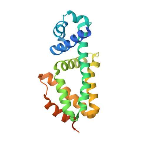 | |
UniProt | |||||
Find proteins for I6X658 (Mycobacterium tuberculosis (strain ATCC 25618 / H37Rv)) Explore I6X658 Go to UniProtKB: I6X658 | |||||
Entity Groups | |||||
| Sequence Clusters | 30% Identity50% Identity70% Identity90% Identity95% Identity100% Identity | ||||
| UniProt Group | I6X658 | ||||
Sequence AnnotationsExpand | |||||
| |||||
| Length ( Å ) | Angle ( ˚ ) |
|---|---|
| a = 78.699 | α = 90 |
| b = 118.866 | β = 90 |
| c = 42.137 | γ = 90 |
| Software Name | Purpose |
|---|---|
| SCALEPACK | data scaling |
| PHENIX | refinement |
| PDB_EXTRACT | data extraction |
| ADSC | data collection |
| HKL-2000 | data reduction |
| PHASER | phasing |
RCSB PDB (citation) is hosted by
RCSB PDB is a member of the

