3T6A

Structure of the C-terminal domain of BCAR3


Experimental Data Snapshot

  • Method: X-RAY DIFFRACTION
  • Resolution: 2.40 Å
  • R-Value Free: 0.244 
  • R-Value Work: 0.174 
  • R-Value Observed: 0.177 

wwPDB Validation   3D Report Full Report


Ligand Structure Quality Assessment 


This is version 1.2 of the entry. See complete history


Literature

NSP-Cas protein structures reveal a promiscuous interaction module in cell signaling.

Mace, P.D.Wallez, Y.Dobaczewska, M.K.Lee, J.J.Robinson, H.Pasquale, E.B.Riedl, S.J.

(2011) Nat Struct Mol Biol 18: 1381-1387

  • DOI: https://doi.org/10.1038/nsmb.2152
  • Primary Citation of Related Structures:  
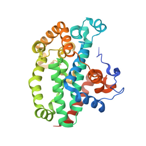
    3T6A, 3T6G

  • PubMed Abstract: 

    Members of the novel SH2-containing protein (NSP) and Crk-associated substrate (Cas) protein families form multidomain signaling platforms that mediate cell migration and invasion through a collection of distinct signaling motifs. Members of each family interact via their respective C-terminal domains, but the mechanism of this association has remained enigmatic. Here we present the crystal structures of the C-terminal domain from the NSP protein BCAR3 and the complex of NSP3 with p130Cas. BCAR3 adopts the Cdc25-homology fold of Ras GTPase exchange factors, but it has a 'closed' conformation incapable of enzymatic activity. The structure of the NSP3-p130Cas complex reveals that this closed conformation is instrumental for interaction of NSP proteins with a focal adhesion-targeting domain present in Cas proteins. This enzyme-to-adaptor conversion enables high-affinity, yet promiscuous, interactions between NSP and Cas proteins and represents an unprecedented mechanistic paradigm linking cellular signaling networks.


  • Organizational Affiliation

    Program of Apoptosis and Cell Death Research, Cancer Center, Sanford-Burnham Medical Research Institute, La Jolla, California, USA.


Macromolecules
Find similar proteins by:  (by identity cutoff)  |  3D Structure
Entity ID: 1
MoleculeChains Sequence LengthOrganismDetailsImage
Breast cancer anti-estrogen resistance protein 3
A, B, C, D
333Homo sapiensMutation(s): 1 
Gene Names: BCAR3NSP2SH2D3BUNQ271/PRO308
UniProt & NIH Common Fund Data Resources
Find proteins for O75815 (Homo sapiens)
Explore O75815 
Go to UniProtKB:  O75815
PHAROS:  O75815
GTEx:  ENSG00000137936 
Entity Groups  
Sequence Clusters30% Identity50% Identity70% Identity90% Identity95% Identity100% Identity
UniProt GroupO75815
Sequence Annotations
Expand
  • Reference Sequence
Small Molecules
Ligands 2 Unique
IDChains Name / Formula / InChI Key2D Diagram3D Interactions
POG
Query on POG

Download Ideal Coordinates CCD File 
E [auth A],
F [auth B]
(20S)-2,5,8,11,14,17-HEXAMETHYL-3,6,9,12,15,18-HEXAOXAHENICOSANE-1,20-DIOL
C21 H44 O8
OWRNLGZKEZSHGO-QAWAPDBNSA-N
UNX
Query on UNX

Download Ideal Coordinates CCD File 
G [auth D]
H [auth D]
I [auth D]
J [auth D]
K [auth D]
G [auth D],
H [auth D],
I [auth D],
J [auth D],
K [auth D],
L [auth D],
M [auth D],
N [auth D]
UNKNOWN ATOM OR ION
X
Experimental Data & Validation

Experimental Data

  • Method: X-RAY DIFFRACTION
  • Resolution: 2.40 Å
  • R-Value Free: 0.244 
  • R-Value Work: 0.174 
  • R-Value Observed: 0.177 
  • Space Group: P 21 21 21
Unit Cell:
Length ( Å )Angle ( ˚ )
a = 50.233α = 90
b = 151.889β = 90
c = 196.503γ = 90
Software Package:
Software NamePurpose
CrystalCleardata collection
PHASERphasing
PHENIXrefinement
XDSdata reduction
SCALAdata scaling

Structure Validation

View Full Validation Report


Ligand Structure Quality Assessment 


Entry History 

Deposition Data

Revision History  (Full details and data files)

  • Version 1.0: 2011-11-23
    Type: Initial release
  • Version 1.1: 2011-12-28
    Changes: Database references
  • Version 1.2: 2024-02-28
    Changes: Data collection, Database references, Derived calculations, Structure summary