3T4N

Structure of the regulatory fragment of Saccharomyces cerevisiae AMPK in complex with ADP


Experimental Data Snapshot

  • Method: X-RAY DIFFRACTION
  • Resolution: 2.30 Å
  • R-Value Free: 0.268 
  • R-Value Work: 0.221 
  • R-Value Observed: 0.223 

wwPDB Validation   3D Report Full Report


Ligand Structure Quality Assessment 


This is version 1.2 of the entry. See complete history


Literature

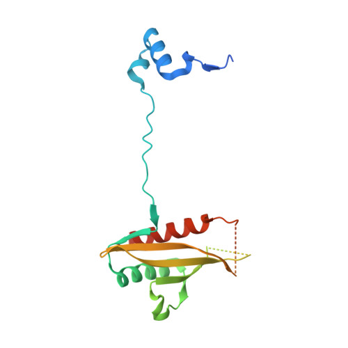
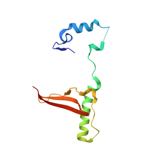
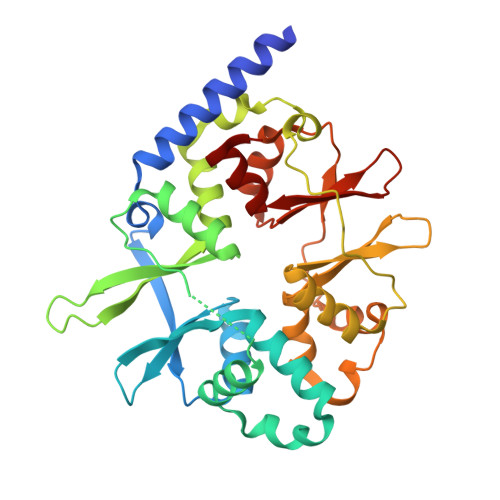
ADP Regulates SNF1, the Saccharomyces cerevisiae Homolog of AMP-Activated Protein Kinase.

Mayer, F.V.Heath, R.Underwood, E.Sanders, M.J.Carmena, D.McCartney, R.R.Leiper, F.C.Xiao, B.Jing, C.Walker, P.A.Haire, L.F.Ogrodowicz, R.Martin, S.R.Schmidt, M.C.Gamblin, S.J.Carling, D.

(2011) Cell Metab 14: 707-714

  • DOI: https://doi.org/10.1016/j.cmet.2011.09.009
  • Primary Citation of Related Structures:  
    3T4N, 3TDH, 3TE5

  • PubMed Abstract: 

    The SNF1 protein kinase complex plays an essential role in regulating gene expression in response to the level of extracellular glucose in budding yeast. SNF1 shares structural and functional similarities with mammalian AMP-activated protein kinase. Both kinases are activated by phosphorylation on a threonine residue within the activation loop segment of the catalytic subunit. Here we show that ADP is the long-sought metabolite that activates SNF1 in response to glucose limitation by protecting the enzyme against dephosphorylation by Glc7, its physiologically relevant protein phosphatase. We also show that the regulatory subunit of SNF1 has two ADP binding sites. The tighter site binds AMP, ADP, and ATP competitively with NADH, whereas the weaker site does not bind NADH, but is responsible for mediating the protective effect of ADP on dephosphorylation. Mutagenesis experiments suggest that the general mechanism by which ADP protects against dephosphorylation is strongly conserved between SNF1 and AMPK.


  • Organizational Affiliation

    MRC Clinical Sciences Centre, Cellular Stress Group, Hammersmith Hospital Campus, Imperial College, DuCane Road, London, UK.


Macromolecules
Find similar proteins by:  (by identity cutoff)  |  3D Structure
Entity ID: 1
MoleculeChains Sequence LengthOrganismDetailsImage
Carbon catabolite-derepressing protein kinase179Saccharomyces cerevisiae S288CMutation(s): 0 
Gene Names: SNF1CAT1CCR1GLC2PAS14YDR477WD8035.20
EC: 2.7.11.1
UniProt
Find proteins for P06782 (Saccharomyces cerevisiae (strain ATCC 204508 / S288c))
Explore P06782 
Go to UniProtKB:  P06782
Entity Groups  
Sequence Clusters30% Identity50% Identity70% Identity90% Identity95% Identity100% Identity
UniProt GroupP06782
Sequence Annotations
Expand
  • Reference Sequence
Find similar proteins by:  (by identity cutoff)  |  3D Structure
Entity ID: 2
MoleculeChains Sequence LengthOrganismDetailsImage
SNF1 protein kinase subunit beta-2113Saccharomyces cerevisiae S288CMutation(s): 0 
Gene Names: SIP2SPM2YGL208WG1155
UniProt
Find proteins for P34164 (Saccharomyces cerevisiae (strain ATCC 204508 / S288c))
Explore P34164 
Go to UniProtKB:  P34164
Entity Groups  
Sequence Clusters30% Identity50% Identity70% Identity90% Identity95% Identity100% Identity
UniProt GroupP34164
Sequence Annotations
Expand
  • Reference Sequence
Find similar proteins by:  (by identity cutoff)  |  3D Structure
Entity ID: 3
MoleculeChains Sequence LengthOrganismDetailsImage
Nuclear protein SNF4323Saccharomyces cerevisiae S288CMutation(s): 0 
Gene Names: SNF4CAT3YGL115W
UniProt
Find proteins for P12904 (Saccharomyces cerevisiae (strain ATCC 204508 / S288c))
Explore P12904 
Go to UniProtKB:  P12904
Entity Groups  
Sequence Clusters30% Identity50% Identity70% Identity90% Identity95% Identity100% Identity
UniProt GroupP12904
Sequence Annotations
Expand
  • Reference Sequence
Small Molecules
Ligands 1 Unique
IDChains Name / Formula / InChI Key2D Diagram3D Interactions
ADP
Query on ADP

Download Ideal Coordinates CCD File 
D [auth C]ADENOSINE-5'-DIPHOSPHATE
C10 H15 N5 O10 P2
XTWYTFMLZFPYCI-KQYNXXCUSA-N
Binding Affinity Annotations 
IDSourceBinding Affinity
ADP Binding MOAD:  3T4N Kd: 7.20e+4 (nM) from 1 assay(s)
PDBBind:  3T4N Kd: 7.20e+4 (nM) from 1 assay(s)
Experimental Data & Validation

Experimental Data

  • Method: X-RAY DIFFRACTION
  • Resolution: 2.30 Å
  • R-Value Free: 0.268 
  • R-Value Work: 0.221 
  • R-Value Observed: 0.223 
  • Space Group: C 2 2 21
Unit Cell:
Length ( Å )Angle ( ˚ )
a = 88.599α = 90
b = 251.214β = 90
c = 79.589γ = 90
Software Package:
Software NamePurpose
HKL-2000data collection
PHASERphasing
REFMACrefinement
DENZOdata reduction
SCALEPACKdata scaling

Structure Validation

View Full Validation Report


Ligand Structure Quality Assessment 


Entry History 

Revision History  (Full details and data files)

  • Version 1.0: 2011-11-09
    Type: Initial release
  • Version 1.1: 2011-11-23
    Changes: Database references
  • Version 1.2: 2024-02-28
    Changes: Data collection, Database references, Derived calculations