3SZG

Crystal structure of C176A glutamine-dependent NAD+ synthetase from M. tuberculosis bound to AMP/PPi and NaAD+


Experimental Data Snapshot

  • Method: X-RAY DIFFRACTION
  • Resolution: 2.25 Å
  • R-Value Free: 0.229 
  • R-Value Work: 0.192 
  • R-Value Observed: 0.194 

wwPDB Validation   3D Report Full Report


Ligand Structure Quality Assessment 


This is version 2.1 of the entry. See complete history


Literature

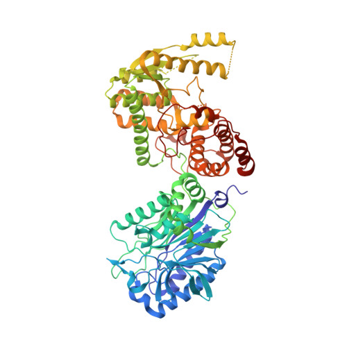
Regulation of the intersubunit ammonia tunnel in Mycobacterium tuberculosis glutamine-dependent NAD+ synthetase.

Chuenchor, W.Doukov, T.I.Resto, M.Chang, A.Gerratana, B.

(2012) Biochem J 443: 417-426

  • DOI: https://doi.org/10.1042/BJ20112210
  • Primary Citation of Related Structures:  
    3SDB, 3SEQ, 3SEZ, 3SYT, 3SZG

  • PubMed Abstract: 

    Glutamine-dependent NAD+ synthetase is an essential enzyme and a validated drug target in Mycobacterium tuberculosis (mtuNadE). It catalyses the ATP-dependent formation of NAD+ from NaAD+ (nicotinic acid-adenine dinucleotide) at the synthetase active site and glutamine hydrolysis at the glutaminase active site. An ammonia tunnel 40 Å (1 Å=0.1 nm) long allows transfer of ammonia from one active site to the other. The enzyme displays stringent kinetic synergism; however, its regulatory mechanism is unclear. In the present paper, we report the structures of the inactive glutaminase C176A variant in an apo form and in three synthetase-ligand complexes with substrates (NaAD+/ATP), substrate analogue {NaAD+/AMP-CPP (adenosine 5'-[α,β-methylene]triphosphate)} and intermediate analogues (NaAD+/AMP/PPi), as well as the structure of wild-type mtuNadE in a product complex (NAD+/AMP/PPi/glutamate). This series of structures provides snapshots of the ammonia tunnel during the catalytic cycle supported also by kinetics and mutagenesis studies. Three major constriction sites are observed in the tunnel: (i) at the entrance near the glutaminase active site; (ii) in the middle of the tunnel; and (iii) at the end near the synthetase active site. Variation in the number and radius of the tunnel constrictions is apparent in the crystal structures and is related to ligand binding at the synthetase domain. These results provide new insight into the regulation of ammonia transport in the intermolecular tunnel of mtuNadE.


  • Organizational Affiliation

    Department of Chemistry and Biochemistry, University of Maryland, College Park, MD 20742-2021, USA.


Macromolecules
Find similar proteins by:  (by identity cutoff)  |  3D Structure
Entity ID: 1
MoleculeChains Sequence LengthOrganismDetailsImage
Glutamine-dependent NAD(+) synthetase
A, B, C, D
680Mycobacterium tuberculosisMutation(s): 1 
Gene Names: MT2513MTCY428.08nadERv2438c
EC: 6.3.5.1
UniProt
Find proteins for P9WJJ3 (Mycobacterium tuberculosis (strain ATCC 25618 / H37Rv))
Explore P9WJJ3 
Go to UniProtKB:  P9WJJ3
Entity Groups  
Sequence Clusters30% Identity50% Identity70% Identity90% Identity95% Identity100% Identity
UniProt GroupP9WJJ3
Sequence Annotations
Expand
  • Reference Sequence
Small Molecules
Ligands 4 Unique
IDChains Name / Formula / InChI Key2D Diagram3D Interactions
DND
Query on DND

Download Ideal Coordinates CCD File 
E [auth A],
H [auth B],
L [auth C],
P [auth D]
NICOTINIC ACID ADENINE DINUCLEOTIDE
C21 H27 N6 O15 P2
SENPVEZBRZQVST-HISDBWNOSA-O
AMP
Query on AMP

Download Ideal Coordinates CCD File 
F [auth A],
I [auth B],
M [auth C],
Q [auth D]
ADENOSINE MONOPHOSPHATE
C10 H14 N5 O7 P
UDMBCSSLTHHNCD-KQYNXXCUSA-N
POP
Query on POP

Download Ideal Coordinates CCD File 
G [auth A],
J [auth B],
N [auth C],
R [auth D]
PYROPHOSPHATE 2-
H2 O7 P2
XPPKVPWEQAFLFU-UHFFFAOYSA-L
GOL
Query on GOL

Download Ideal Coordinates CCD File 
K [auth B],
O [auth C],
S [auth D]
GLYCEROL
C3 H8 O3
PEDCQBHIVMGVHV-UHFFFAOYSA-N
Experimental Data & Validation

Experimental Data

  • Method: X-RAY DIFFRACTION
  • Resolution: 2.25 Å
  • R-Value Free: 0.229 
  • R-Value Work: 0.192 
  • R-Value Observed: 0.194 
  • Space Group: P 41 21 2
Unit Cell:
Length ( Å )Angle ( ˚ )
a = 177.94α = 90
b = 177.94β = 90
c = 214.94γ = 90
Software Package:
Software NamePurpose
HKL-2000data collection
PHASERphasing
PHENIXrefinement
XDSdata reduction
XDSdata scaling

Structure Validation

View Full Validation Report


Ligand Structure Quality Assessment 


Entry History 

Deposition Data

Revision History  (Full details and data files)

  • Version 1.0: 2012-04-11
    Type: Initial release
  • Version 1.1: 2017-11-08
    Changes: Refinement description
  • Version 2.0: 2019-10-23
    Changes: Atomic model, Data collection, Database references, Derived calculations, Non-polymer description, Structure summary
  • Version 2.1: 2023-09-13
    Changes: Data collection, Database references, Derived calculations, Refinement description