Structural Analysis of Histo-Blood Group Antigen Binding Specificity in a Norovirus GII.4 Epidemic Variant: Implications for Epochal Evolution.
Shanker, S., Choi, J.M., Sankaran, B., Atmar, R.L., Estes, M.K., Prasad, B.V.(2011) J Virol 85: 8635-8645
- PubMed: 21715503
- DOI: https://doi.org/10.1128/JVI.00848-11
- Primary Citation of Related Structures:
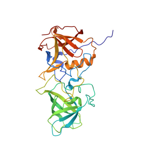
3SEJ, 3SJP, 3SKB, 3SLD, 3SLN - PubMed Abstract:
Susceptibility to norovirus (NoV), a major pathogen of epidemic gastroenteritis, is associated with histo-blood group antigens (HBGAs), which are also cell attachment factors for this virus. GII.4 NoV strains are predominantly associated with worldwide NoV epidemics with a periodic emergence of new variants. The sequence variations in the surface-exposed P domain of the capsid protein resulting in differential HBGA binding patterns and antigenicity are suggested to drive GII.4 epochal evolution. To understand how temporal sequence variations affect the P domain structure and contribute to epochal evolution, we determined the P domain structure of a 2004 variant with ABH and secretor Lewis HBGAs and compared it with the previously determined structure of a 1996 variant. We show that temporal sequence variations do not affect the binding of monofucosyl ABH HBGAs but that they can modulate the binding strength of difucosyl Lewis HBGAs and thus could contribute to epochal evolution by the potentiated targeting of new variants to Lewis-positive, secretor-positive individuals. The temporal variations also result in significant differences in the electrostatic landscapes, likely reflecting antigenic variations. The proximity of some of these changes to the HBGA binding sites suggests the possibility of a coordinated interplay between antigenicity and HBGA binding in epochal evolution. From the observation that the regions involved in the formation of the HBGA binding sites can be conformationally flexible, we suggest a plausible mechanism for how norovirus disassociates from salivary mucin-linked HBGA before reassociating with HBGAs linked to intestinal epithelial cells during its passage through the gastrointestinal tract.
Organizational Affiliation:
Verna Marrs Mclean Department of Biochemistry and Molecular Biology, Baylor College of Medicine, One Baylor Plaza, Houston, TX 77030, USA.