3SE7

ancient VanA


Experimental Data Snapshot

  • Method: X-RAY DIFFRACTION
  • Resolution: 3.07 Å
  • R-Value Free: 0.263 
  • R-Value Work: 0.193 
  • R-Value Observed: 0.197 

wwPDB Validation   3D Report Full Report


Ligand Structure Quality Assessment 


This is version 1.2 of the entry. See complete history


Literature

Antibiotic resistance is ancient.

D'Costa, V.M.King, C.E.Kalan, L.Morar, M.Sung, W.W.Schwarz, C.Froese, D.Zazula, G.Calmels, F.Debruyne, R.Golding, G.B.Poinar, H.N.Wright, G.D.

(2011) Nature 477: 457-461

  • DOI: https://doi.org/10.1038/nature10388
  • Primary Citation of Related Structures:  
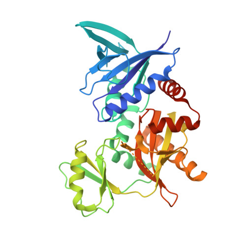
    3SE7

  • PubMed Abstract: 

    The discovery of antibiotics more than 70 years ago initiated a period of drug innovation and implementation in human and animal health and agriculture. These discoveries were tempered in all cases by the emergence of resistant microbes. This history has been interpreted to mean that antibiotic resistance in pathogenic bacteria is a modern phenomenon; this view is reinforced by the fact that collections of microbes that predate the antibiotic era are highly susceptible to antibiotics. Here we report targeted metagenomic analyses of rigorously authenticated ancient DNA from 30,000-year-old Beringian permafrost sediments and the identification of a highly diverse collection of genes encoding resistance to β-lactam, tetracycline and glycopeptide antibiotics. Structure and function studies on the complete vancomycin resistance element VanA confirmed its similarity to modern variants. These results show conclusively that antibiotic resistance is a natural phenomenon that predates the modern selective pressure of clinical antibiotic use.


  • Organizational Affiliation

    Michael G. DeGroote Institute for Infectious Disease Research, McMaster University, Hamilton, Ontario, Canada, L8N 3Z5.


Macromolecules
Find similar proteins by:  (by identity cutoff)  |  3D Structure
Entity ID: 1
MoleculeChains Sequence LengthOrganismDetailsImage
VanA
A, B, C, D, E
A, B, C, D, E, F
346N/AMutation(s): 0 
Gene Names: VanA
Entity Groups  
Sequence Clusters30% Identity50% Identity70% Identity90% Identity95% Identity100% Identity
Sequence Annotations
Expand
  • Reference Sequence
Small Molecules
Ligands 2 Unique
IDChains Name / Formula / InChI Key2D Diagram3D Interactions
ATP
Query on ATP

Download Ideal Coordinates CCD File 
G [auth A]
J [auth B]
M [auth C]
P [auth D]
S [auth E]
G [auth A],
J [auth B],
M [auth C],
P [auth D],
S [auth E],
V [auth F]
ADENOSINE-5'-TRIPHOSPHATE
C10 H16 N5 O13 P3
ZKHQWZAMYRWXGA-KQYNXXCUSA-N
MG
Query on MG

Download Ideal Coordinates CCD File 
H [auth A]
I [auth A]
K [auth B]
L [auth B]
N [auth C]
H [auth A],
I [auth A],
K [auth B],
L [auth B],
N [auth C],
O [auth C],
Q [auth D],
R [auth D],
T [auth E],
U [auth E],
W [auth F],
X [auth F]
MAGNESIUM ION
Mg
JLVVSXFLKOJNIY-UHFFFAOYSA-N
Experimental Data & Validation

Experimental Data

  • Method: X-RAY DIFFRACTION
  • Resolution: 3.07 Å
  • R-Value Free: 0.263 
  • R-Value Work: 0.193 
  • R-Value Observed: 0.197 
  • Space Group: P 21 21 21
Unit Cell:
Length ( Å )Angle ( ˚ )
a = 84.133α = 90
b = 136.032β = 90
c = 178.09γ = 90
Software Package:
Software NamePurpose
HKL-2000data collection
SOLVEphasing
REFMACrefinement
HKL-2000data reduction
HKL-2000data scaling

Structure Validation

View Full Validation Report


Ligand Structure Quality Assessment 


Entry History 

Deposition Data

Revision History  (Full details and data files)

  • Version 1.0: 2011-09-14
    Type: Initial release
  • Version 1.1: 2011-10-19
    Changes: Database references
  • Version 1.2: 2024-02-28
    Changes: Data collection, Database references, Derived calculations