3SAN

Crystal structure of influenza A virus neuraminidase N5 complexed with Zanamivir


Experimental Data Snapshot

  • Method: X-RAY DIFFRACTION
  • Resolution: 1.60 Å
  • R-Value Free: 0.157 
  • R-Value Work: 0.120 
  • R-Value Observed: 0.122 

wwPDB Validation   3D Report Full Report


This is version 2.1 of the entry. See complete history


Literature

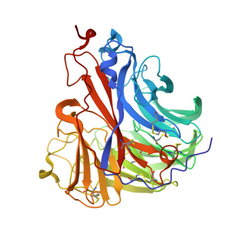
Influenza a virus n5 neuraminidase has an extended 150-cavity

Wang, M.Y.Qi, J.X.Liu, Y.Vavricka, C.J.Wu, Y.Li, Q.Gao, G.F.

(2011) J Virol 85: 8431-8435

  • DOI: https://doi.org/10.1128/JVI.00638-11
  • Primary Citation of Related Structures:  
    3SAL, 3SAN

  • PubMed Abstract: 

    There are 9 serotypes of neuraminidase (NA) from influenza A virus (N1 to N9), which are classified into two groups based on primary sequences (groups 1 and 2). The structural hallmark of the two groups is the presence or absence of an extra 150-cavity (formed by the 150-loop) in the active site. Thus far, structures of NAs from 6 out of the 9 serotypes have been solved. Here, we solved the N5 structure, the last unknown structure group 1 serotype with a unique Asn147 residue in its 150-loop, demonstrating that it has an extended 150-cavity that closes upon inhibitor binding.


  • Organizational Affiliation

    CAS Key Laboratory of Pathogenic Microbiology and Immunology, Institute of Microbiology, Chinese Academy of Sciences, Chaoyang District, Beijing 100101, The People's Republic of China. gaof@im.ac.cn


Macromolecules
Find similar proteins by:  (by identity cutoff)  |  3D Structure
Entity ID: 1
MoleculeChains Sequence LengthOrganismDetailsImage
Neuraminidase
A, B
395Influenza A virus (A/duck/Alberta/60/1976(H12N5))Mutation(s): 0 
Gene Names: NA
EC: 3.2.1.18
UniProt
Find proteins for A1ILL9 (Influenza A virus (strain A/Duck/Alberta/60/1976 H12N5))
Explore A1ILL9 
Go to UniProtKB:  A1ILL9
Entity Groups  
Sequence Clusters30% Identity50% Identity70% Identity90% Identity95% Identity100% Identity
UniProt GroupA1ILL9
Sequence Annotations
Expand
  • Reference Sequence
Oligosaccharides

Help

Entity ID: 2
MoleculeChains Length2D Diagram Glycosylation3D Interactions
2-acetamido-2-deoxy-beta-D-glucopyranose-(1-4)-2-acetamido-2-deoxy-beta-D-glucopyranose
C, D
2N-Glycosylation
Glycosylation Resources
GlyTouCan:  G42666HT
GlyCosmos:  G42666HT
GlyGen:  G42666HT
Small Molecules
Ligands 4 Unique
IDChains Name / Formula / InChI Key2D Diagram3D Interactions
ZMR
Query on ZMR

Download Ideal Coordinates CCD File 
G [auth A],
L [auth B]
ZANAMIVIR
C12 H20 N4 O7
ARAIBEBZBOPLMB-UFGQHTETSA-N
NAG
Query on NAG

Download Ideal Coordinates CCD File 
F [auth A],
K [auth B]
2-acetamido-2-deoxy-beta-D-glucopyranose
C8 H15 N O6
OVRNDRQMDRJTHS-FMDGEEDCSA-N
GOL
Query on GOL

Download Ideal Coordinates CCD File 
H [auth A],
I [auth A],
M [auth B],
N [auth B]
GLYCEROL
C3 H8 O3
PEDCQBHIVMGVHV-UHFFFAOYSA-N
CA
Query on CA

Download Ideal Coordinates CCD File 
E [auth A],
J [auth B]
CALCIUM ION
Ca
BHPQYMZQTOCNFJ-UHFFFAOYSA-N
Experimental Data & Validation

Experimental Data

  • Method: X-RAY DIFFRACTION
  • Resolution: 1.60 Å
  • R-Value Free: 0.157 
  • R-Value Work: 0.120 
  • R-Value Observed: 0.122 
  • Space Group: P 4
Unit Cell:
Length ( Å )Angle ( ˚ )
a = 112.11α = 90
b = 112.11β = 90
c = 66.767γ = 90
Software Package:
Software NamePurpose
ADSCdata collection
PHASERphasing
PHENIXrefinement
HKL-2000data reduction
HKL-2000data scaling

Structure Validation

View Full Validation Report


Entry History 

Deposition Data

Revision History  (Full details and data files)

  • Version 1.0: 2011-08-10
    Type: Initial release
  • Version 2.0: 2020-07-29
    Type: Remediation
    Reason: Carbohydrate remediation
    Changes: Atomic model, Data collection, Derived calculations, Structure summary
  • Version 2.1: 2023-11-01
    Changes: Data collection, Database references, Refinement description, Structure summary