


Crystal structure of ribose-5-phosphate isomerase B RpiB from Giardia lamblia
Edwards, T.E., Abendroth, J., Seattle Structural Genomics Center for Infectious Disease (SSGCID)To be published.
Experimental Data Snapshot
wwPDB Validation 3D Report Full Report

Entity ID: 1 | |||||
|---|---|---|---|---|---|
| Molecule | Chains | Sequence Length | Organism | Details | Image |
| Ribose 5-phosphate isomerase | 166 | Giardia lamblia ATCC 50803 | Mutation(s): 0 Gene Names: GL50803_27614 EC: 5.3.1.6 | 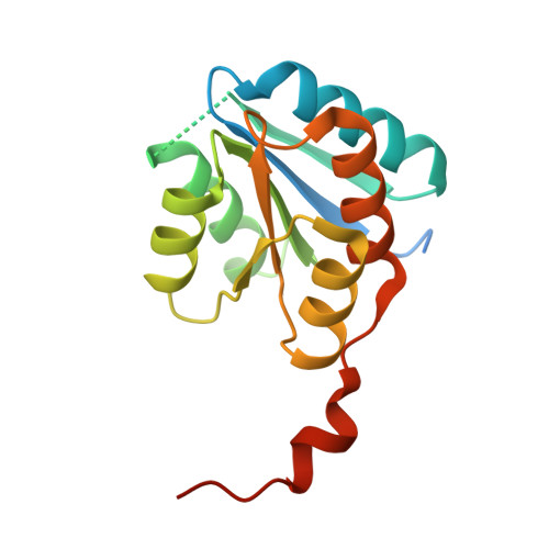 | |
UniProt | |||||
Find proteins for A8B2K2 (Giardia intestinalis (strain ATCC 50803 / WB clone C6)) Explore A8B2K2 Go to UniProtKB: A8B2K2 | |||||
Entity Groups | |||||
| Sequence Clusters | 30% Identity50% Identity70% Identity90% Identity95% Identity100% Identity | ||||
| UniProt Group | A8B2K2 | ||||
Sequence AnnotationsExpand | |||||
| |||||
| Ligands 1 Unique | |||||
|---|---|---|---|---|---|
| ID | Chains | Name / Formula / InChI Key | 2D Diagram | 3D Interactions | |
| SO4 Query on SO4 | C [auth A], D [auth A], E [auth B], F [auth B], G [auth B] | SULFATE ION O4 S QAOWNCQODCNURD-UHFFFAOYSA-L |  | ||
| Length ( Å ) | Angle ( ˚ ) |
|---|---|
| a = 89.2 | α = 90 |
| b = 89.2 | β = 90 |
| c = 68.24 | γ = 120 |
| Software Name | Purpose |
|---|---|
| XSCALE | data scaling |
| MOLREP | phasing |
| REFMAC | refinement |
| PDB_EXTRACT | data extraction |
| XDS | data reduction |
RCSB PDB (citation) is hosted by
RCSB PDB is a member of the

