3RUX

Crystal structure of biotin-protein ligase BirA from Mycobacterium tuberculosis in complex with an acylsulfamide bisubstrate inhibitor


Experimental Data Snapshot

  • Method: X-RAY DIFFRACTION
  • Resolution: 1.70 Å
  • R-Value Free: 0.216 
  • R-Value Work: 0.190 
  • R-Value Observed: 0.192 

wwPDB Validation   3D Report Full Report


Ligand Structure Quality Assessment 


This is version 1.1 of the entry. See complete history


Literature

Bisubstrate Adenylation Inhibitors of Biotin Protein Ligase from Mycobacterium tuberculosis.

Duckworth, B.P.Geders, T.W.Tiwari, D.Boshoff, H.I.Sibbald, P.A.Barry, C.E.Schnappinger, D.Finzel, B.C.Aldrich, C.C.

(2011) Chem Biol 18: 1432-1441

  • DOI: https://doi.org/10.1016/j.chembiol.2011.08.013
  • Primary Citation of Related Structures:  
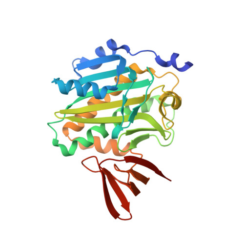
    3RUX

  • PubMed Abstract: 

    The mycobacterial biotin protein ligase (MtBPL) globally regulates lipid metabolism in Mtb through the posttranslational biotinylation of acyl coenzyme A carboxylases involved in lipid biosynthesis that catalyze the first step in fatty acid biosynthesis and pyruvate coenzyme A carboxylase, a gluconeogenic enzyme vital for lipid catabolism. Here we describe the design, development, and evaluation of a rationally designed bisubstrate inhibitor of MtBPL. This inhibitor displays potent subnanomolar enzyme inhibition and antitubercular activity against multidrug resistant and extensively drug resistant Mtb strains. We show that the inhibitor decreases in vivo protein biotinylation of key enzymes involved in fatty acid biosynthesis and that the antibacterial activity is MtBPL dependent. Additionally, the gene encoding BPL was found to be essential in M. smegmatis. Finally, the X-ray cocrystal structure of inhibitor bound MtBPL was solved providing detailed insight for further structure-activity analysis. Collectively, these data suggest that MtBPL is a promising target for further antitubercular therapeutic development.


  • Organizational Affiliation

    Center for Drug Design, University of Minnesota, Minneapolis, MN 55455, USA.


Macromolecules
Find similar proteins by:  (by identity cutoff)  |  3D Structure
Entity ID: 1
MoleculeChains Sequence LengthOrganismDetailsImage
BirA bifunctional protein
A, B
270Mycobacterium tuberculosisMutation(s): 0 
Gene Names: birAMT3379Rv3279c
EC: 6.3.4.15
UniProt
Find proteins for P96884 (Mycobacterium tuberculosis (strain CDC 1551 / Oshkosh))
Explore P96884 
Go to UniProtKB:  P96884
Entity Groups  
Sequence Clusters30% Identity50% Identity70% Identity90% Identity95% Identity100% Identity
UniProt GroupP96884
Sequence Annotations
Expand
  • Reference Sequence
Small Molecules
Ligands 1 Unique
IDChains Name / Formula / InChI Key2D Diagram3D Interactions
BS5
Query on BS5

Download Ideal Coordinates CCD File 
C [auth A],
D [auth B]
5'-deoxy-5'-[({5-[(3aS,4S,6aR)-2-oxohexahydro-1H-thieno[3,4-d]imidazol-4-yl]pentanoyl}sulfamoyl)amino]adenosine
C20 H29 N9 O7 S2
RSZBCKJLQVSFKT-RHCAYAJFSA-N
Binding Affinity Annotations 
IDSourceBinding Affinity
BS5 Binding MOAD:  3RUX Kd: 0.53 (nM) from 1 assay(s)
BindingDB:  3RUX Kd: 0.87 (nM) from 1 assay(s)
PDBBind:  3RUX Kd: 0.53 (nM) from 1 assay(s)
Experimental Data & Validation

Experimental Data

  • Method: X-RAY DIFFRACTION
  • Resolution: 1.70 Å
  • R-Value Free: 0.216 
  • R-Value Work: 0.190 
  • R-Value Observed: 0.192 
  • Space Group: P 21 21 21
Unit Cell:
Length ( Å )Angle ( ˚ )
a = 63.882α = 90
b = 68.802β = 90
c = 115.755γ = 90
Software Package:
Software NamePurpose
Blu-Icedata collection
PHASERphasing
PHENIXrefinement
d*TREKdata reduction
d*TREKdata scaling

Structure Validation

View Full Validation Report


Ligand Structure Quality Assessment 


Entry History 

Deposition Data

Revision History  (Full details and data files)

  • Version 1.0: 2011-12-14
    Type: Initial release
  • Version 1.1: 2024-02-28
    Changes: Data collection, Database references, Derived calculations