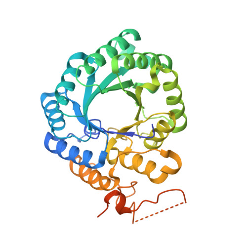
Removal of the C-terminal regulatory domain of alpha-isopropylmalate synthase disrupts functional substrate binding
Huisman, F.H.A., Koon, N., Bulloch, E.M.M., Baker, H.M., Baker, E.N., Squire, C.J., Parker, E.J.(2012) Biochemistry 51: 2289-2297
- PubMed: 22352945
- DOI: https://doi.org/10.1021/bi201717j
- Primary Citation of Related Structures:
3HPX, 3RMJ, 3U6W - PubMed Abstract:
α-Isopropylmalate synthase (α-IPMS) catalyzes the metal-dependent aldol reaction between α-ketoisovalerate (α-KIV) and acetyl-coenzyme A (AcCoA) to give α-isopropylmalate (α-IPM). This reaction is the first committed step in the biosynthesis of leucine in bacteria. α-IPMS is homodimeric, with monomers consisting of (β/α)(8) barrel catalytic domains fused to a C-terminal regulatory domain, responsible for binding leucine and providing feedback regulation for leucine biosynthesis. In these studies, we demonstrate that removal of the regulatory domain from the α-IPMS enzymes of both Neisseria meningitidis (NmeIPMS) and Mycobacterium tuberculosis (MtuIPMS) results in enzymes that are unable to catalyze the formation of α-IPM, although truncated NmeIPMS was still able to slowly hydrolyze AcCoA. The lack of catalytic activity of these truncation variants was confirmed by complementation studies with Escherichia coli cells lacking the α-IPMS gene, where transformation with the plasmids encoding the truncated α-IPMS enzymes was not able to rescue α-IPMS activity. X-ray crystal structures of both truncation variants reveal that both proteins are dimeric and that the catalytic sites of the proteins are intact, although the divalent metal ion that is thought to be responsible for activating substrate α-KIV is displaced slightly relative to its position in the substrate-bound, wild-type structure. Isothermal titration calorimetry and WaterLOGSY nuclear magnetic resonance experiments demonstrate that although these truncation variants are not able to catalyze the reaction between α-KIV and AcCoA, they are still able to bind the substrate α-KIV. It is proposed that the regulatory domain is crucial for ensuring protein dynamics necessary for competent catalysis.
Organizational Affiliation:
Biomolecular Interaction Centre and Department of Chemistry, University of Canterbury, Christchurch, New Zealand.