3REN

CPF_2247, a novel alpha-amylase from Clostridium perfringens


Experimental Data Snapshot

  • Method: X-RAY DIFFRACTION
  • Resolution: 2.00 Å
  • R-Value Free: 0.224 
  • R-Value Work: 0.169 
  • R-Value Observed: 0.172 

wwPDB Validation   3D Report Full Report


This is version 1.2 of the entry. See complete history


Literature

Structural analysis of CPF_2247, a novel alpha-amylase from Clostridium perfringens.

Ficko-Blean, E.Stuart, C.P.Boraston, A.B.

(2011) Proteins 79: 2771-2777

  • DOI: https://doi.org/10.1002/prot.23116

  • PubMed Abstract: 

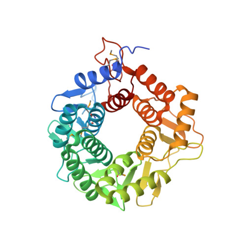
    CPF_2247 from Clostridium perfringens ATCC 13124 was identified as a putative carbohydrate-active enzyme by its low sequence identity to endo-β-1,4-glucanases belonging to family 8 of the glycoside hydrolase classification. The X-ray crystal structure of CPF_2247 determined to 2.0 Å resolution by single-wavelength anomalous dispersion using seleno-methionine-substituted protein revealed an (α/α)(6) barrel fold. A large cleft on the surface of the protein contains residues that are structurally conserved with key elements of the catalytic machinery in clan GH-M glycoside hydrolases. Assessment of CPF_2247 as a carbohydrate-active enzyme disclosed α-glucanase activity on amylose, glycogen, and malto-oligosaccharides.


  • Organizational Affiliation

    Department of Biochemistry & Microbiology, University of Victoria, Victoria, British Columbia, Canada.


Macromolecules
Find similar proteins by:  (by identity cutoff)  |  3D Structure
Entity ID: 1
MoleculeChains Sequence LengthOrganismDetailsImage
Glycosyl hydrolase, family 8
A, B
350Clostridium perfringensMutation(s): 0 
Gene Names: CPF_2247
UniProt
Find proteins for A0A0H2YP60 (Clostridium perfringens (strain ATCC 13124 / DSM 756 / JCM 1290 / NCIMB 6125 / NCTC 8237 / Type A))
Explore A0A0H2YP60 
Go to UniProtKB:  A0A0H2YP60
Entity Groups  
Sequence Clusters30% Identity50% Identity70% Identity90% Identity95% Identity100% Identity
UniProt GroupA0A0H2YP60
Sequence Annotations
Expand
  • Reference Sequence
Small Molecules
Ligands 3 Unique
IDChains Name / Formula / InChI Key2D Diagram3D Interactions
SO4
Query on SO4

Download Ideal Coordinates CCD File 
E [auth A],
N [auth B],
O [auth B]
SULFATE ION
O4 S
QAOWNCQODCNURD-UHFFFAOYSA-L
EDO
Query on EDO

Download Ideal Coordinates CCD File 
F [auth A]
G [auth A]
H [auth A]
I [auth A]
J [auth A]
F [auth A],
G [auth A],
H [auth A],
I [auth A],
J [auth A],
K [auth A],
L [auth A],
P [auth B],
Q [auth B],
R [auth B],
S [auth B],
T [auth B],
U [auth B]
1,2-ETHANEDIOL
C2 H6 O2
LYCAIKOWRPUZTN-UHFFFAOYSA-N
MG
Query on MG

Download Ideal Coordinates CCD File 
C [auth A],
D [auth A],
M [auth B]
MAGNESIUM ION
Mg
JLVVSXFLKOJNIY-UHFFFAOYSA-N
Modified Residues  1 Unique
IDChains TypeFormula2D DiagramParent
MSE
Query on MSE
A, B
L-PEPTIDE LINKINGC5 H11 N O2 SeMET
Experimental Data & Validation

Experimental Data

  • Method: X-RAY DIFFRACTION
  • Resolution: 2.00 Å
  • R-Value Free: 0.224 
  • R-Value Work: 0.169 
  • R-Value Observed: 0.172 
  • Space Group: P 21 21 21
Unit Cell:
Length ( Å )Angle ( ˚ )
a = 65.35α = 90
b = 75.06β = 90
c = 158.54γ = 90
Software Package:
Software NamePurpose
REFMACrefinement

Structure Validation

View Full Validation Report


Entry History 

Deposition Data

Revision History  (Full details and data files)

  • Version 1.0: 2011-05-18
    Type: Initial release
  • Version 1.1: 2011-07-13
    Changes: Version format compliance
  • Version 1.2: 2013-09-04
    Changes: Database references