3QW2

L-myo-inositol 1-phosphate synthase from Archaeoglobus mutant N255A


Experimental Data Snapshot

  • Method: X-RAY DIFFRACTION
  • Resolution: 2.59 Å
  • R-Value Free: 0.267 
  • R-Value Work: 0.185 
  • R-Value Observed: 0.189 

wwPDB Validation   3D Report Full Report


Ligand Structure Quality Assessment 


This is version 1.4 of the entry. See complete history


Literature

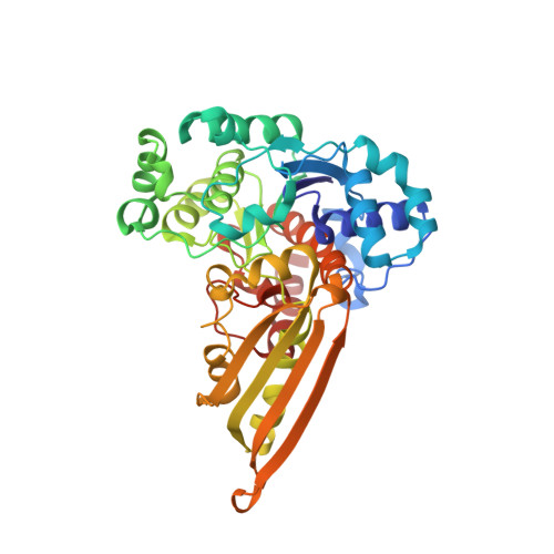
Crystal structure of a trapped catalytic intermediate suggests that forced atomic proximity drives the catalysis of mIPS.

Neelon, K.Roberts, M.F.Stec, B.

(2011) Biophys J 101: 2816-2824

  • DOI: https://doi.org/10.1016/j.bpj.2011.10.038
  • Primary Citation of Related Structures:  
    3QVS, 3QVT, 3QVW, 3QVX, 3QW2

  • PubMed Abstract: 

    1-L-myo-inositol-phosphate synthase (mIPS) catalyzes the first step of the unique, de novo pathway of inositol biosynthesis. However, details about the complex mIPS catalytic mechanism, which requires oxidation, enolization, intramolecular aldol cyclization, and reduction, are not fully known. To gain further insight into this mechanism, we determined the crystal structure of the wild-type mIPS from Archaeoglobus fulgidus at 1.7 Å, as well as the crystal structures of three active-site mutants. Additionally, we obtained the structure of mIPS with a trapped 5-keto-glucose-6-phosphate intermediate at 2 Å resolution by a novel (to our knowledge) process of activating the crystal at high temperature. A comparison of all of the crystal structures of mIPS described in this work suggests a novel type of catalytic mechanism that relies on the forced atomic proximity of functional groups. The lysine cluster is contained in a small volume in the active site, where random motions of these side chains are responsible for the progress of the complex multistep reaction as well as for the low rate of catalysis. The mechanism requires that functional groups of Lys-274, Lys-278, Lys-306, and Lys-367 assume differential roles in the protonation/deprotonation steps that must occur during the mIPS reaction. This mechanism is supported by the complete loss of activity of the enzyme caused by the Leu-257 mutation to Ala that releases the lysine containment.


  • Organizational Affiliation

    Department of Chemistry, Boston College, Chestnut Hill, Massachusetts, USA.


Macromolecules
Find similar proteins by:  (by identity cutoff)  |  3D Structure
Entity ID: 1
MoleculeChains Sequence LengthOrganismDetailsImage
Myo-inositol-1-phosphate synthase (Ino1)
A, B, C, D
392Archaeoglobus fulgidusMutation(s): 1 
Gene Names: AF1794AF_1794
EC: 5.5.1.4
UniProt
Find proteins for O28480 (Archaeoglobus fulgidus (strain ATCC 49558 / DSM 4304 / JCM 9628 / NBRC 100126 / VC-16))
Explore O28480 
Go to UniProtKB:  O28480
Entity Groups  
Sequence Clusters30% Identity50% Identity70% Identity90% Identity95% Identity100% Identity
UniProt GroupO28480
Sequence Annotations
Expand
  • Reference Sequence
Small Molecules
Ligands 5 Unique
IDChains Name / Formula / InChI Key2D Diagram3D Interactions
NAD
Query on NAD

Download Ideal Coordinates CCD File 
F [auth A],
K [auth B],
P [auth C],
V [auth D]
NICOTINAMIDE-ADENINE-DINUCLEOTIDE
C21 H27 N7 O14 P2
BAWFJGJZGIEFAR-NNYOXOHSSA-N
SO4
Query on SO4

Download Ideal Coordinates CCD File 
H [auth A]
M [auth B]
N [auth B]
Q [auth C]
R [auth C]
H [auth A],
M [auth B],
N [auth B],
Q [auth C],
R [auth C],
S [auth C],
W [auth D],
X [auth D]
SULFATE ION
O4 S
QAOWNCQODCNURD-UHFFFAOYSA-L
PO4
Query on PO4

Download Ideal Coordinates CCD File 
E [auth A],
J [auth B],
O [auth C],
U [auth D]
PHOSPHATE ION
O4 P
NBIIXXVUZAFLBC-UHFFFAOYSA-K
GOL
Query on GOL

Download Ideal Coordinates CCD File 
G [auth A],
L [auth B]
GLYCEROL
C3 H8 O3
PEDCQBHIVMGVHV-UHFFFAOYSA-N
K
Query on K

Download Ideal Coordinates CCD File 
I [auth A],
T [auth C],
Y [auth D]
POTASSIUM ION
K
NPYPAHLBTDXSSS-UHFFFAOYSA-N
Experimental Data & Validation

Experimental Data

  • Method: X-RAY DIFFRACTION
  • Resolution: 2.59 Å
  • R-Value Free: 0.267 
  • R-Value Work: 0.185 
  • R-Value Observed: 0.189 
  • Space Group: P 1 21 1
Unit Cell:
Length ( Å )Angle ( ˚ )
a = 91.512α = 90
b = 88.036β = 94.91
c = 103.727γ = 90
Software Package:
Software NamePurpose
REFMACrefinement
CNSrefinement
CrystalCleardata collection
CrystalCleardata reduction
HKL-2000data scaling
CNSphasing

Structure Validation

View Full Validation Report


Ligand Structure Quality Assessment 


Entry History 

Deposition Data

Revision History  (Full details and data files)

  • Version 1.0: 2012-01-11
    Type: Initial release
  • Version 1.1: 2018-03-07
    Changes: Data collection
  • Version 1.2: 2019-07-17
    Changes: Data collection, Refinement description
  • Version 1.3: 2022-12-07
    Changes: Database references, Derived calculations
  • Version 1.4: 2023-09-20
    Changes: Data collection, Refinement description