3QBU

Crystal structure of putative peptidoglycan deactelyase (HP0310) from Helicobacter pylori


Experimental Data Snapshot

  • Method: X-RAY DIFFRACTION
  • Resolution: 2.57 Å
  • R-Value Free: 0.235 
  • R-Value Work: 0.204 
  • R-Value Observed: 0.206 

wwPDB Validation   3D Report Full Report


This is version 1.3 of the entry. See complete history


Literature

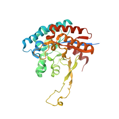
The Structure of Helicobacter pylori HP0310 Reveals an Atypical Peptidoglycan Deacetylase.

Shaik, M.M.Cendron, L.Percudani, R.Zanotti, G.

(2011) PLoS One 6: e19207-e19207

  • DOI: https://doi.org/10.1371/journal.pone.0019207
  • Primary Citation of Related Structures:  
    3QBU

  • PubMed Abstract: 

    Peptidoglycan deacetlyase (HP0310, HpPgdA) from the gram-negative pathogen Helicobacter pylori, has been indicated as the enzyme responsible for a peptidoglycan modification that counteracts the host immune response. HpPgdA has been cloned, purified and expressed in good yield in E. coli. It has been crystallized, its structure determined and activity tests in vitro performed. The enzyme, which belongs to the polysaccharide deacetylases protein family, is a homo-tetramer. The four polypeptide chains, each folded into a single domain characterized by a non-canonical TIM-barrel fold, are arranged around a four-fold symmetry axis. The active site, one per monomer, contains a heavy ion coordinated in a way similar to other deacetylases. However, the enzyme showed no in vitro activity on the typical polysaccharide substrates of peptidoglycan deacetylases. In striking contrast with the known peptidoglycan deacetylases, HpPgdA does not exhibit a solvent-accessible polysaccharide binding groove, suggesting that the enzyme binds a small molecule at the active site.


  • Organizational Affiliation

    Department of Biological Chemistry, University of Padua, Padua, Italy.


Macromolecules
Find similar proteins by:  (by identity cutoff)  |  3D Structure
Entity ID: 1
MoleculeChains Sequence LengthOrganismDetailsImage
Putative uncharacterized protein
A, B, C, D
326Helicobacter pylori G27Mutation(s): 0 
Gene Names: hp0310HPG27_289
UniProt
Find proteins for B5ZA76 (Helicobacter pylori (strain G27))
Explore B5ZA76 
Go to UniProtKB:  B5ZA76
Entity Groups  
Sequence Clusters30% Identity50% Identity70% Identity90% Identity95% Identity100% Identity
UniProt GroupB5ZA76
Sequence Annotations
Expand
  • Reference Sequence
Experimental Data & Validation

Experimental Data

  • Method: X-RAY DIFFRACTION
  • Resolution: 2.57 Å
  • R-Value Free: 0.235 
  • R-Value Work: 0.204 
  • R-Value Observed: 0.206 
  • Space Group: C 2 2 21
Unit Cell:
Length ( Å )Angle ( ˚ )
a = 154.56α = 90
b = 154.73β = 90
c = 158.68γ = 90
Software Package:
Software NamePurpose
ADSCdata collection
PHASERphasing
PHENIXrefinement
MOSFLMdata reduction
SCALAdata scaling

Structure Validation

View Full Validation Report


Entry History 

Deposition Data

Revision History  (Full details and data files)

  • Version 1.0: 2011-05-25
    Type: Initial release
  • Version 1.1: 2011-07-13
    Changes: Version format compliance
  • Version 1.2: 2013-11-20
    Changes: Structure summary
  • Version 1.3: 2023-09-13
    Changes: Data collection, Database references, Derived calculations, Refinement description