Conformational basis for substrate recognition and regulation of catalytic activity in Staphylococcus aureus nucleoside di-phosphate kinase.
Srivastava, S.K., Rajasree, K., Gopal, B.(2011) Biochim Biophys Acta
- PubMed: 21745603
- DOI: https://doi.org/10.1016/j.bbapap.2011.06.008
- Primary Citation of Related Structures:
3Q83, 3Q86, 3Q89, 3Q8U, 3Q8V, 3Q8Y - PubMed Abstract:
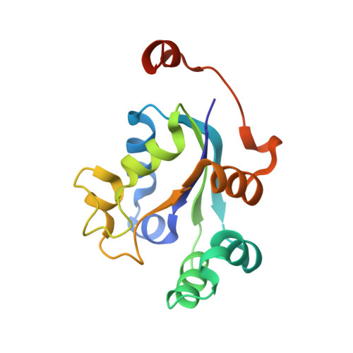
Nucleoside diphosphate kinases (NDK) are characterized by high catalytic turnover rates and diverse substrate specificity. These features make this enzyme an effective activator of a pro-drug-an application that has been actively pursued for a variety of therapeutic strategies. The catalytic mechanism of this enzyme is governed by a conserved histidine that coordinates a magnesium ion at the active site. Despite substantial structural and biochemical information on NDK, the mechanistic feature of the phospho-transfer that leads to auto-phosphorylation remains unclear. While the role of the histidine residue is well documented, the other active site residues, in particular the conserved serine remains poorly characterized. Studies on some homologues suggest no role for the serine residue at the active site, while others suggest a crucial role for this serine in the regulation and quaternary association of this enzyme in some species. Here we report the biochemical features of the Staphylococcus aureus NDK and the mutant enzymes. We also describe the crystal structures of the apo-NDK, as a transition state mimic with vanadate and in complex with different nucleotide substrates. These structures formed the basis for molecular dynamics simulations to understand the broad substrate specificity of this enzyme and the role of active site residues in the phospho-transfer mechanism and oligomerization. Put together, these data suggest that concerted changes in the conformation of specific residues facilitate the stabilization of nucleotide complexes thereby enabling the steps involved in the ping-pong reaction mechanism without large changes to the overall structure of this enzyme.
Organizational Affiliation:
Indian Institute of Science, Bangalore, India. sandeep@mbu.iisc.ernet.in