3Q2P

Reduced sweetness of a monellin (MNEI) mutant results from increased protein flexibility and disruption of a distant poly-(L-proline) II helix


Experimental Data Snapshot

  • Method: X-RAY DIFFRACTION
  • Resolution: 2.34 Å
  • R-Value Free: 0.245 
  • R-Value Work: 0.184 
  • R-Value Observed: 0.187 

wwPDB Validation   3D Report Full Report


This is version 1.3 of the entry. See complete history


Literature

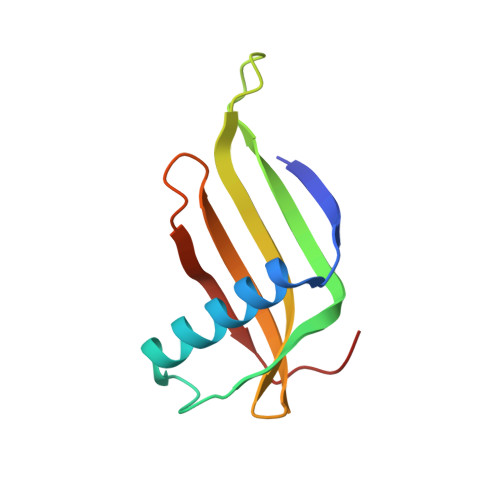
Reduced Sweetness of a Monellin (MNEI) Mutant Results from Increased Protein Flexibility and Disruption of a Distant Poly-(L-Proline) II Helix.

Templeton, C.M.Ostovar Pour, S.Hobbs, J.R.Blanch, E.W.Munger, S.D.Conn, G.L.

(2011) Chem Senses 36: 425-434

  • DOI: https://doi.org/10.1093/chemse/bjr007
  • Primary Citation of Related Structures:  
    3PXM, 3PYJ, 3Q2P

  • PubMed Abstract: 

    Monellin is a highly potent sweet-tasting protein but relatively little is known about how it interacts with the sweet taste receptor. We determined X-ray crystal structures of 3 single-chain monellin (MNEI) proteins with alterations at 2 core residues (G16A, V37A, and G16A/V37A) that induce 2- to 10-fold reductions in sweetness relative to the wild-type protein. Surprisingly, no changes were observed in the global protein fold or the positions of surface amino acids important for MNEI sweetness that could explain these differences in protein activity. Differential scanning calorimetry showed that while the thermal stability of each mutant MNEI was reduced, the least sweet mutant, G16A-MNEI, was not the least stable protein. In contrast, solution spectroscopic measurements revealed that changes in protein flexibility and the C-terminal structure correlate directly with protein activity. G16A mutation-induced disorder in the protein core is propagated via changes to hydrophobic interactions that disrupt the formation and/or position of a critical C-terminal poly-(L-proline) II helix. These findings suggest that MNEI interaction with the sweet taste receptor is highly sensitive to the relative positions of key residues across its protein surface and that loss of sweetness in G16A-MNEI may result from an increased entropic cost of binding.


  • Organizational Affiliation

    Department of Biochemistry, Emory University School of Medicine, Atlanta, GA 30322, USA.


Macromolecules
Find similar proteins by:  (by identity cutoff)  |  3D Structure
Entity ID: 1
MoleculeChains Sequence LengthOrganismDetailsImage
Monellin chain B/Monellin chain A chimeric protein
A, B, C, D
97Dioscoreophyllum cumminsiiMutation(s): 2 
UniProt
Find proteins for P02881 (Dioscoreophyllum cumminsii)
Explore P02881 
Go to UniProtKB:  P02881
Find proteins for P02882 (Dioscoreophyllum cumminsii)
Explore P02882 
Go to UniProtKB:  P02882
Entity Groups  
Sequence Clusters30% Identity50% Identity70% Identity90% Identity95% Identity100% Identity
UniProt GroupsP02881P02882
Sequence Annotations
Expand
  • Reference Sequence
Small Molecules
Ligands 1 Unique
IDChains Name / Formula / InChI Key2D Diagram3D Interactions
NA
Query on NA

Download Ideal Coordinates CCD File 
E [auth A]SODIUM ION
Na
FKNQFGJONOIPTF-UHFFFAOYSA-N
Experimental Data & Validation

Experimental Data

  • Method: X-RAY DIFFRACTION
  • Resolution: 2.34 Å
  • R-Value Free: 0.245 
  • R-Value Work: 0.184 
  • R-Value Observed: 0.187 
  • Space Group: P 1 21 1
Unit Cell:
Length ( Å )Angle ( ˚ )
a = 31.4α = 90
b = 144.1β = 90
c = 45.8γ = 90
Software Package:
Software NamePurpose
CrystalCleardata collection
MOLREPphasing
PHENIXrefinement
XDSdata reduction
XSCALEdata scaling

Structure Validation

View Full Validation Report


Entry History 

Deposition Data

Revision History  (Full details and data files)

  • Version 1.0: 2011-04-06
    Type: Initial release
  • Version 1.1: 2011-07-13
    Changes: Version format compliance
  • Version 1.2: 2017-08-02
    Changes: Advisory, Refinement description, Source and taxonomy
  • Version 1.3: 2023-09-13
    Changes: Advisory, Data collection, Database references, Refinement description