3Q2I

Crystal structure of the WlbA dehydrognase from Chromobactrium violaceum in complex with NADH and UDP-GlcNAcA at 1.50 A resolution


Experimental Data Snapshot

  • Method: X-RAY DIFFRACTION
  • Resolution: 1.50 Å
  • R-Value Free: 0.236 
  • R-Value Work: 0.201 
  • R-Value Observed: 0.203 

wwPDB Validation   3D Report Full Report


Ligand Structure Quality Assessment 


This is version 1.3 of the entry. See complete history


Literature

Biochemical and Structural Characterization of WlbA from Bordetella pertussis and Chromobacterium violaceum: Enzymes Required for the Biosynthesis of 2,3-Diacetamido-2,3-dideoxy-d-mannuronic Acid.

Thoden, J.B.Holden, H.M.

(2011) Biochemistry 50: 1483-1491

  • DOI: https://doi.org/10.1021/bi101871f
  • Primary Citation of Related Structures:  
    3Q2I, 3Q2K

  • PubMed Abstract: 

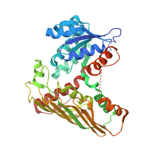
    The unusual sugar 2,3-diacetamido-2,3-dideoxy-d-mannuronic acid, or ManNAc3NAcA, has been observed in the lipopolysaccharides of both pathogenic and nonpathogenic Gram-negative bacteria. It is added to the lipopolysaccharides of these organisms by glycosyltransferases that use as substrates UDP-ManNAc3NAcA. Five enzymes are ultimately required for the biosynthesis of UDP-ManNAc3NAcA starting from UDP-N-acetylglucosamine. The second enzyme in the pathway, encoded by the wlba gene and referred to as WlbA, catalyzes the NAD-dependent oxidation of the C-3' hydroxyl group of the UDP-linked sugar. Here we describe a combined structural and functional investigation of the WlbA enzymes from Bordetella pertussis and Chromobacterium violaceum. For this investigation, ternary structures were determined in the presence of NAD(H) and substrate to 2.13 and 1.5 Å resolution, respectively. Both of the enzymes display octameric quaternary structures with their active sites positioned far apart. The octamers can be envisioned as tetramers of dimers. Kinetic studies demonstrate that the reaction mechanisms for these enzymes are sequential and that they do not require α-ketoglutarate for activity. These results are in sharp contrast to those recently reported for the WlbA enzymes from Pseudomonas aeruginosa and Thermus thermophilus, which function via ping-pong mechanisms that involve α-ketoglutarate. Taken together, the results reported here demonstrate that there are two distinct families of WlbA enzymes, which differ with respect to amino acid sequences, quaternary structures, active site architectures, and kinetic mechanisms.


  • Organizational Affiliation

    Department of Biochemistry, University of Wisconsin, Madison, Wisconsin 53706, United States.


Macromolecules
Find similar proteins by:  (by identity cutoff)  |  3D Structure
Entity ID: 1
MoleculeChains Sequence LengthOrganismDetailsImage
dehydrogenase354Chromobacterium violaceumMutation(s): 0 
Gene Names: CV_4127wlbA
UniProt
Find proteins for Q7NQL0 (Chromobacterium violaceum (strain ATCC 12472 / DSM 30191 / JCM 1249 / NBRC 12614 / NCIMB 9131 / NCTC 9757))
Explore Q7NQL0 
Go to UniProtKB:  Q7NQL0
Entity Groups  
Sequence Clusters30% Identity50% Identity70% Identity90% Identity95% Identity100% Identity
UniProt GroupQ7NQL0
Sequence Annotations
Expand
  • Reference Sequence
Small Molecules
Ligands 4 Unique
IDChains Name / Formula / InChI Key2D Diagram3D Interactions
NAI
Query on NAI

Download Ideal Coordinates CCD File 
B [auth A]1,4-DIHYDRONICOTINAMIDE ADENINE DINUCLEOTIDE
C21 H29 N7 O14 P2
BOPGDPNILDQYTO-NNYOXOHSSA-N
HP7
Query on HP7

Download Ideal Coordinates CCD File 
C [auth A](2S,3S,4R,5R,6R)-5-acetamido-6-[[[(2R,3S,4R,5R)-5-(2,4-dioxopyrimidin-1-yl)-3,4-dihydroxy-oxolan-2-yl]methoxy-hydroxy-phosphoryl]oxy-hydroxy-phosphoryl]oxy-3,4-dihydroxy-oxane-2-carboxylic acid
C17 H25 N3 O18 P2
DZOGQXKQLXAPND-HHKCBAECSA-N
CL
Query on CL

Download Ideal Coordinates CCD File 
D [auth A]CHLORIDE ION
Cl
VEXZGXHMUGYJMC-UHFFFAOYSA-M
NA
Query on NA

Download Ideal Coordinates CCD File 
E [auth A]SODIUM ION
Na
FKNQFGJONOIPTF-UHFFFAOYSA-N
Experimental Data & Validation

Experimental Data

  • Method: X-RAY DIFFRACTION
  • Resolution: 1.50 Å
  • R-Value Free: 0.236 
  • R-Value Work: 0.201 
  • R-Value Observed: 0.203 
  • Space Group: I 4 2 2
Unit Cell:
Length ( Å )Angle ( ˚ )
a = 106.711α = 90
b = 106.711β = 90
c = 143.356γ = 90
Software Package:
Software NamePurpose
HKL-3000data collection
SOLVEphasing
REFMACrefinement
HKL-3000data reduction
HKL-3000data scaling

Structure Validation

View Full Validation Report


Ligand Structure Quality Assessment 


Entry History 

Deposition Data

Revision History  (Full details and data files)

  • Version 1.0: 2011-01-19
    Type: Initial release
  • Version 1.1: 2011-07-13
    Changes: Version format compliance
  • Version 1.2: 2013-11-06
    Changes: Non-polymer description
  • Version 1.3: 2024-02-21
    Changes: Data collection, Database references, Derived calculations