3Q0B

Crystal structure of SUVH5 SRA- fully methylated CG DNA complex in space group P42212


Experimental Data Snapshot

  • Method: X-RAY DIFFRACTION
  • Resolution: 2.20 Å
  • R-Value Free: 0.252 
  • R-Value Work: 0.217 
  • R-Value Observed: 0.218 

wwPDB Validation   3D Report Full Report


This is version 1.1 of the entry. See complete history


Literature

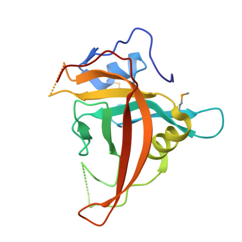
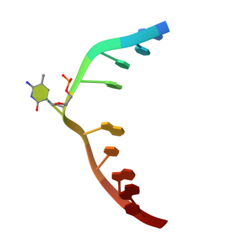
A dual flip-out mechanism for 5mC recognition by the Arabidopsis SUVH5 SRA domain and its impact on DNA methylation and H3K9 dimethylation in vivo.

Rajakumara, E.Law, J.A.Simanshu, D.K.Voigt, P.Johnson, L.M.Reinberg, D.Patel, D.J.Jacobsen, S.E.

(2011) Genes Dev 25: 137-152

  • DOI: https://doi.org/10.1101/gad.1980311
  • Primary Citation of Related Structures:  
    3Q0B, 3Q0C, 3Q0D, 3Q0F

  • PubMed Abstract: 

    Cytosine DNA methylation is evolutionarily ancient, and in eukaryotes this epigenetic modification is associated with gene silencing. Proteins with SRA (SET- or RING-associated) methyl-binding domains are required for the establishment and/or maintenance of DNA methylation in both plants and mammals. The 5-methyl-cytosine (5mC)-binding specificity of several SRA domains have been characterized, and each one has a preference for DNA methylation in different sequence contexts. Here we demonstrate through mobility shift assays and calorimetric measurements that the SU(VAR)3-9 HOMOLOG 5 (SUVH5) SRA domain differs from other SRA domains in that it can bind methylated DNA in all contexts to similar extents. Crystal structures of the SUVH5 SRA domain bound to 5mC-containing DNA in either the fully or hemimethylated CG context or the methylated CHH context revealed a dual flip-out mechanism where both the 5mC and a base (5mC, C, or G, respectively) from the partner strand are simultaneously extruded from the DNA duplex and positioned within binding pockets of individual SRA domains. Our structure-based in vivo studies suggest that a functional SUVH5 SRA domain is required for both DNA methylation and accumulation of the H3K9 dimethyl modification in vivo, suggesting a role for the SRA domain in recruitment of SUVH5 to genomic loci.


  • Organizational Affiliation

    Structural Biology Program, Memorial Sloan-Kettering Cancer Center, New York, New York 10065, USA.


Macromolecules

Find similar proteins by:  (by identity cutoff)  |  3D Structure
Entity ID: 1
MoleculeChains Sequence LengthOrganismDetailsImage
Histone-lysine N-methyltransferase, H3 lysine-9 specific SUVH5A [auth X]167Arabidopsis thalianaMutation(s): 0 
Gene Names: At2g35160SDG9SET9SUVH5T4C15.17
EC: 2.1.1.43
UniProt
Find proteins for O82175 (Arabidopsis thaliana)
Explore O82175 
Go to UniProtKB:  O82175
Entity Groups  
Sequence Clusters30% Identity50% Identity70% Identity90% Identity95% Identity100% Identity
UniProt GroupO82175
Sequence Annotations
Expand
  • Reference Sequence

Find similar nucleic acids by:  Sequence   |   3D Structure  

Entity ID: 2
MoleculeChains LengthOrganismImage
DNA (5'-D(*AP*CP*TP*AP*(5CM)P*GP*TP*AP*GP*T)-3')B [auth A]10N/A
Sequence Annotations
Expand
  • Reference Sequence
Small Molecules
Modified Residues  1 Unique
IDChains TypeFormula2D DiagramParent
MSE
Query on MSE
A [auth X]L-PEPTIDE LINKINGC5 H11 N O2 SeMET
Experimental Data & Validation

Experimental Data

  • Method: X-RAY DIFFRACTION
  • Resolution: 2.20 Å
  • R-Value Free: 0.252 
  • R-Value Work: 0.217 
  • R-Value Observed: 0.218 
  • Space Group: P 42 21 2
Unit Cell:
Length ( Å )Angle ( ˚ )
a = 76.566α = 90
b = 76.566β = 90
c = 73.904γ = 90
Software Package:
Software NamePurpose
ADSCdata collection
SHARPphasing
PHENIXrefinement
HKL-2000data reduction
HKL-2000data scaling

Structure Validation

View Full Validation Report


Entry History 

Deposition Data

Revision History  (Full details and data files)

  • Version 1.0: 2011-02-02
    Type: Initial release
  • Version 1.1: 2011-07-13
    Changes: Version format compliance